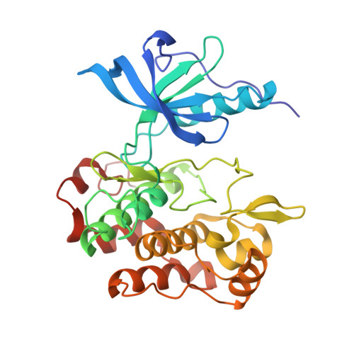
5HHW

Crystal structure of insulin receptor kinase domain in complex with cis-(R)-7-(3-(azetidin-1-ylmethyl)cyclobutyl)-5-(3-((tetrahydro-2H-pyran-2-yl)methoxy)phenyl)-7H-pyrrolo[2,3-d]pyrimidin-4-amine.


Experimental Data Snapshot

  • Method: X-RAY DIFFRACTION
  • Resolution: 1.79 Å
  • R-Value Free: 0.198 
  • R-Value Work: 0.171 
  • R-Value Observed: 0.172 

wwPDB Validation   3D Report Full Report


Ligand Structure Quality Assessment 


This is version 1.1 of the entry. See complete history


Literature

Identification of a 5-[3-phenyl-(2-cyclic-ether)-methylether]-4-aminopyrrolo[2,3-d]pyrimidine series of IGF-1R inhibitors.

Stauffer, F.Cowan-Jacob, S.W.Scheufler, C.Furet, P.

(2016) Bioorg Med Chem Lett 26: 2065-2067

  • DOI: https://doi.org/10.1016/j.bmcl.2016.02.074
  • Primary Citation of Related Structures:  
    5HHW, 5HZN

  • PubMed Abstract: 

    We report structure-guided modifications of the benzyloxy substituent of the Insulin-like Growth Factor-1 Receptor (IGF-1R) inhibitor NVP-AEW541. This chemical group has been shown to confer selectivity against other protein kinases but at the expense of a metabolism liability. X-ray crystallography has revealed that the benzyloxy moiety interacts with a lysine cation of the IGF-1R kinase domain via its ether function and its aromatic π-system and is nicely embedded in an induced hydrophobic pocket. We show that 1,4-diethers displaying an adequate hydrophobic and constrained shape are advantageous benzyloxy replacements. A single digit nanomolar inhibitor (compound 20, IC50=8.9 nM) was identified following this approach.


  • Organizational Affiliation

    Novartis Institutes for Biomedical Research, Basel, Postfach, 4002 Basel, Switzerland. Electronic address: frederic.stauffer@novartis.com.


Macromolecules
Find similar proteins by:  (by identity cutoff)  |  3D Structure
Entity ID: 1
MoleculeChains Sequence LengthOrganismDetailsImage
Insulin receptor307Homo sapiensMutation(s): 2 
Gene Names: INSR
EC: 2.7.10.1
UniProt & NIH Common Fund Data Resources
Find proteins for P06213 (Homo sapiens)
Explore P06213 
Go to UniProtKB:  P06213
PHAROS:  P06213
GTEx:  ENSG00000171105 
Entity Groups  
Sequence Clusters30% Identity50% Identity70% Identity90% Identity95% Identity100% Identity
UniProt GroupP06213
Sequence Annotations
Expand
  • Reference Sequence
Small Molecules
Ligands 2 Unique
IDChains Name / Formula / InChI Key2D Diagram3D Interactions
60O
Query on 60O

Download Ideal Coordinates CCD File 
B [auth A]7-[3-(azetidin-1-ylmethyl)cyclobutyl]-5-[3-[[(2~{R})-oxan-2-yl]methoxy]phenyl]pyrrolo[2,3-d]pyrimidin-4-amine
C26 H33 N5 O2
UTHVKVUADCCITP-KAGYGMCKSA-N
EDO
Query on EDO

Download Ideal Coordinates CCD File 
C [auth A]1,2-ETHANEDIOL
C2 H6 O2
LYCAIKOWRPUZTN-UHFFFAOYSA-N
Experimental Data & Validation

Experimental Data

  • Method: X-RAY DIFFRACTION
  • Resolution: 1.79 Å
  • R-Value Free: 0.198 
  • R-Value Work: 0.171 
  • R-Value Observed: 0.172 
  • Space Group: P 21 21 21
Unit Cell:
Length ( Å )Angle ( ˚ )
a = 57.92α = 90
b = 69.16β = 90
c = 89.86γ = 90
Software Package:
Software NamePurpose
BUSTERrefinement
XDSdata reduction
XSCALEdata scaling
PHASERphasing

Structure Validation

View Full Validation Report


Ligand Structure Quality Assessment 


Entry History 

Deposition Data

Revision History  (Full details and data files)

  • Version 1.0: 2016-04-13
    Type: Initial release
  • Version 1.1: 2018-01-24
    Changes: Source and taxonomy