5HH5

Crystal structure of B3 metallo-beta-lactamase L1 complexed with a phosphonate-based inhibitor


Experimental Data Snapshot

  • Method: X-RAY DIFFRACTION
  • Resolution: 1.80 Å
  • R-Value Free: 0.198 
  • R-Value Work: 0.163 
  • R-Value Observed: 0.165 

wwPDB Validation   3D Report Full Report


Ligand Structure Quality Assessment 


This is version 1.5 of the entry. See complete history


Literature

Structural and Kinetic Studies of the Potent Inhibition of Metallo-beta-lactamases by 6-Phosphonomethylpyridine-2-carboxylates.

Hinchliffe, P.Tanner, C.A.Krismanich, A.P.Labbe, G.Goodfellow, V.J.Marrone, L.Desoky, A.Y.Calvopina, K.Whittle, E.E.Zeng, F.Avison, M.B.Bols, N.C.Siemann, S.Spencer, J.Dmitrienko, G.I.

(2018) Biochemistry 57: 1880-1892

  • DOI: https://doi.org/10.1021/acs.biochem.7b01299
  • Primary Citation of Related Structures:  
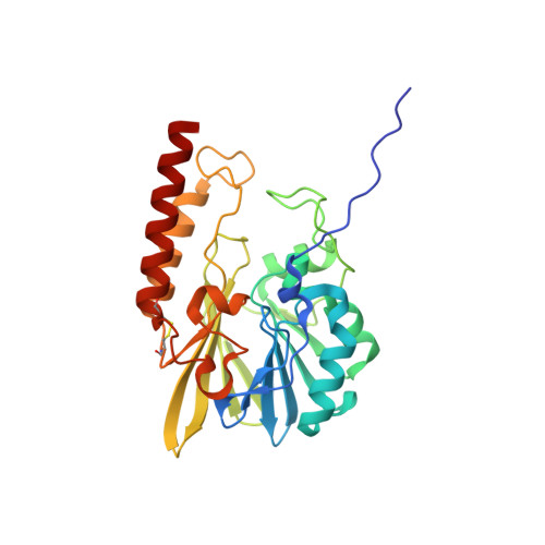
    5HH4, 5HH5, 5HH6

  • PubMed Abstract: 

    There are currently no clinically available inhibitors of metallo-β-lactamases (MBLs), enzymes that hydrolyze β-lactam antibiotics and confer resistance to Gram-negative bacteria. Here we present 6-phosphonomethylpyridine-2-carboxylates (PMPCs) as potent inhibitors of subclass B1 (IMP-1, VIM-2, and NDM-1) and B3 (L1) MBLs. Inhibition followed a competitive, slow-binding model without an isomerization step (IC 50 values of 0.3-7.2 μM; K i values of 0.03-1.5 μM). Minimum inhibitory concentration assays demonstrated potentiation of β-lactam (Meropenem) activity against MBL-producing bacteria, including clinical isolates, at concentrations at which eukaryotic cells remain viable. Crystal structures revealed unprecedented modes of binding of inhibitor to B1 (IMP-1) and B3 (L1) MBLs. In IMP-1, binding does not replace the nucleophilic hydroxide, and the PMPC carboxylate and pyridine nitrogen interact closely (2.3 and 2.7 Å, respectively) with the Zn2 ion of the binuclear metal site. The phosphonate group makes limited interactions but is 2.6 Å from the nucleophilic hydroxide. Furthermore, the presence of a water molecule interacting with the PMPC phosphonate and pyridine N-C2 π-bond, as well as the nucleophilic hydroxide, suggests that the PMPC binds to the MBL active site as its hydrate. Binding is markedly different in L1, with the phosphonate displacing both Zn2, forming a monozinc enzyme, and the nucleophilic hydroxide, while also making multiple interactions with the protein main chain and Zn1. The carboxylate and pyridine nitrogen interact with Ser221 and -223, respectively (3 Å distance). The potency, low toxicity, cellular activity, and amenability to further modification of PMPCs indicate these and similar phosphonate compounds can be further considered for future MBL inhibitor development.


  • Organizational Affiliation

    School of Cellular & Molecular Medicine , University of Bristol , Bristol BS8 1TD , U.K.


Macromolecules
Find similar proteins by:  (by identity cutoff)  |  3D Structure
Entity ID: 1
MoleculeChains Sequence LengthOrganismDetailsImage
Metallo-beta-lactamase L1271Stenotrophomonas maltophiliaMutation(s): 0 
EC: 3.5.2.6
UniProt
Find proteins for P52700 (Stenotrophomonas maltophilia)
Explore P52700 
Go to UniProtKB:  P52700
Entity Groups  
Sequence Clusters30% Identity50% Identity70% Identity90% Identity95% Identity100% Identity
UniProt GroupP52700
Sequence Annotations
Expand
  • Reference Sequence
Small Molecules
Ligands 4 Unique
IDChains Name / Formula / InChI Key2D Diagram3D Interactions
60M
Query on 60M

Download Ideal Coordinates CCD File 
H [auth A]6-(phosphonomethyl)pyridine-2-carboxylic acid
C7 H8 N O5 P
HIDJRVUSOKZHOS-UHFFFAOYSA-N
SO4
Query on SO4

Download Ideal Coordinates CCD File 
C [auth A],
D [auth A],
E [auth A],
F [auth A]
SULFATE ION
O4 S
QAOWNCQODCNURD-UHFFFAOYSA-L
GOL
Query on GOL

Download Ideal Coordinates CCD File 
G [auth A]GLYCEROL
C3 H8 O3
PEDCQBHIVMGVHV-UHFFFAOYSA-N
ZN
Query on ZN

Download Ideal Coordinates CCD File 
B [auth A]ZINC ION
Zn
PTFCDOFLOPIGGS-UHFFFAOYSA-N
Experimental Data & Validation

Experimental Data

  • Method: X-RAY DIFFRACTION
  • Resolution: 1.80 Å
  • R-Value Free: 0.198 
  • R-Value Work: 0.163 
  • R-Value Observed: 0.165 
  • Space Group: P 64 2 2
Unit Cell:
Length ( Å )Angle ( ˚ )
a = 105.256α = 90
b = 105.256β = 90
c = 98.573γ = 120
Software Package:
Software NamePurpose
PHENIXrefinement
XDSdata reduction
Aimlessdata scaling
PHASERphasing

Structure Validation

View Full Validation Report


Ligand Structure Quality Assessment 


Entry History & Funding Information

Deposition Data


Funding OrganizationLocationGrant Number
Medical Research Council (United Kingdom)United KingdomG1100135
National Institutes of Health/National Institute Of Allergy and Infectious Diseases (NIH/NIAID)United StatesR01AI100560

Revision History  (Full details and data files)

  • Version 1.0: 2017-01-18
    Type: Initial release
  • Version 1.1: 2017-09-13
    Changes: Author supporting evidence
  • Version 1.2: 2019-02-27
    Changes: Data collection, Database references
  • Version 1.3: 2021-04-14
    Changes: Database references, Derived calculations
  • Version 1.4: 2022-03-30
    Changes: Author supporting evidence, Database references, Derived calculations
  • Version 1.5: 2024-01-10
    Changes: Data collection, Refinement description