5HGJ

Structure of integrin alpha1beta1 and alpha2beta1 I-domains explain differential calcium-mediated ligand recognition


Experimental Data Snapshot

  • Method: X-RAY DIFFRACTION
  • Resolution: 1.40 Å
  • R-Value Free: 0.205 
  • R-Value Work: 0.178 
  • R-Value Observed: 0.179 

wwPDB Validation   3D Report Full Report


This is version 1.4 of the entry. See complete history


Literature

Salt-bridge modulates differential calcium-mediated ligand binding to integrin alpha 1- and alpha 2-I domains.

Brown, K.L.Banerjee, S.Feigley, A.Abe, H.Blackwell, T.S.Pozzi, A.Hudson, B.G.Zent, R.

(2018) Sci Rep 8: 2916-2916

  • DOI: https://doi.org/10.1038/s41598-018-21231-1
  • Primary Citation of Related Structures:  
    5HGJ, 5HJ2

  • PubMed Abstract: 

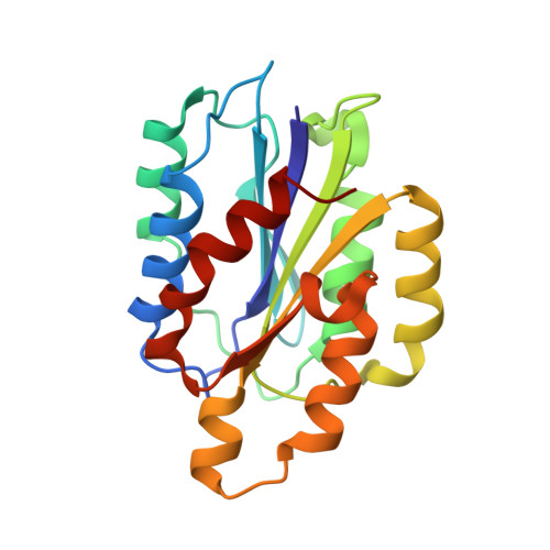
    Integrins are transmembrane cell-extracellular matrix adhesion receptors that impact many cellular functions. A subgroup of integrins contain an inserted (I) domain within the α-subunits (αI) that mediate ligand recognition where function is contingent on binding a divalent cation at the metal ion dependent adhesion site (MIDAS). Ca 2+ is reported to promote α1I but inhibit α2I ligand binding. We co-crystallized individual I-domains with MIDAS-bound Ca 2+ and report structures at 1.4 and 2.15 Å resolution, respectively. Both structures are in the "closed" ligand binding conformation where Ca 2+ induces minimal global structural changes. Comparisons with Mg 2+ -bound structures reveal Mg 2+ and Ca 2+ bind α1I in a manner sufficient to promote ligand binding. In contrast, Ca 2+ is displaced in the α2I domain MIDAS by 1.4 Å relative to Mg 2+ and unable to directly coordinate all MIDAS residues. We identified an E152-R192 salt bridge hypothesized to limit the flexibility of the α2I MIDAS, thus, reducing Ca 2+ binding. A α2I E152A construct resulted in a 10,000-fold increase in Mg 2+ and Ca 2+ binding affinity while increasing binding to collagen ligands 20%. These data indicate the E152-R192 salt bridge is a key distinction in the molecular mechanism of differential ion binding of these two I domains.


  • Organizational Affiliation

    Department of Medicine, Vanderbilt University Medical Center, Nashville, TN, 37232-2372, USA. kyle.l.brown@vanderbilt.edu.


Macromolecules
Find similar proteins by:  (by identity cutoff)  |  3D Structure
Entity ID: 1
MoleculeChains Sequence LengthOrganismDetailsImage
Integrin alpha-1
A, B
195Homo sapiensMutation(s): 0 
Gene Names: ITGA1
UniProt & NIH Common Fund Data Resources
Find proteins for P56199 (Homo sapiens)
Explore P56199 
Go to UniProtKB:  P56199
PHAROS:  P56199
GTEx:  ENSG00000213949 
Entity Groups  
Sequence Clusters30% Identity50% Identity70% Identity90% Identity95% Identity100% Identity
UniProt GroupP56199
Sequence Annotations
Expand
  • Reference Sequence
Experimental Data & Validation

Experimental Data

  • Method: X-RAY DIFFRACTION
  • Resolution: 1.40 Å
  • R-Value Free: 0.205 
  • R-Value Work: 0.178 
  • R-Value Observed: 0.179 
  • Space Group: P 1 21 1
Unit Cell:
Length ( Å )Angle ( ˚ )
a = 37.369α = 90
b = 95.95β = 104
c = 53.078γ = 90
Software Package:
Software NamePurpose
PHENIXrefinement
HKL-2000data reduction
SCALAdata scaling
PHASERphasing

Structure Validation

View Full Validation Report


Entry History & Funding Information

Deposition Data


Funding OrganizationLocationGrant Number
National Institutes of Health/National Heart, Lung, and Blood Institute (NIH/NHLBI)United StatesT32 HL94296-06
National Institutes of Health/National Institute of Diabetes and Digestive and Kidney Disease (NIH/NIDDK)United StatesR01 DK018381

Revision History  (Full details and data files)

  • Version 1.0: 2017-04-12
    Type: Initial release
  • Version 1.1: 2017-09-20
    Changes: Author supporting evidence
  • Version 1.2: 2018-02-28
    Changes: Database references
  • Version 1.3: 2019-12-04
    Changes: Author supporting evidence
  • Version 1.4: 2024-03-06
    Changes: Data collection, Database references, Derived calculations