5H9A

Crystal structure of the Apo form of human cellular retinol binding protein 1


Experimental Data Snapshot

  • Method: X-RAY DIFFRACTION
  • Resolution: 1.38 Å
  • R-Value Free: 0.171 
  • R-Value Work: 0.133 
  • R-Value Observed: 0.135 

wwPDB Validation   3D Report Full Report


This is version 1.5 of the entry. See complete history


Literature

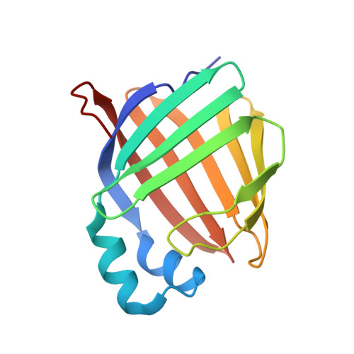
Ligand Binding Induces Conformational Changes in Human Cellular Retinol-binding Protein 1 (CRBP1) Revealed by Atomic Resolution Crystal Structures.

Silvaroli, J.A.Arne, J.M.Chelstowska, S.Kiser, P.D.Banerjee, S.Golczak, M.

(2016) J Biol Chem 291: 8528-8540

  • DOI: https://doi.org/10.1074/jbc.M116.714535
  • Primary Citation of Related Structures:  
    5H8T, 5H9A, 5HA1, 5HBS

  • PubMed Abstract: 

    Important in regulating the uptake, storage, and metabolism of retinoids, cellular retinol-binding protein 1 (CRBP1) is essential for trafficking vitamin A through the cytoplasm. However, the molecular details of ligand uptake and targeted release by CRBP1 remain unclear. Here we report the first structure of CRBP1 in a ligand-free form as well as ultra-high resolution structures of this protein bound to either all-trans-retinol or retinylamine, the latter a therapeutic retinoid that prevents light-induced retinal degeneration. Superpositioning of human apo- and holo-CRBP1 revealed major differences within segments surrounding the entrance to the retinoid-binding site. These included α-helix II and hairpin turns between β-strands βC-βD and βE-βF as well as several side chains, such as Phe-57, Tyr-60, and Ile-77, that change their orientations to accommodate the ligand. Additionally, we mapped hydrogen bond networks inside the retinoid-binding cavity and demonstrated their significance for the ligand affinity. Analyses of the crystallographic B-factors indicated several regions with higher backbone mobility in the apoprotein that became more rigid upon retinoid binding. This conformational flexibility of human apo-CRBP1 facilitates interaction with the ligands, whereas the more rigid holoprotein structure protects the labile retinoid moiety during vitamin A transport. These findings suggest a mechanism of induced fit upon ligand binding by mammalian cellular retinol-binding proteins.


  • Organizational Affiliation

    From the Department of Pharmacology and.


Macromolecules
Find similar proteins by:  (by identity cutoff)  |  3D Structure
Entity ID: 1
MoleculeChains Sequence LengthOrganismDetailsImage
Retinol-binding protein 1140Homo sapiensMutation(s): 0 
Gene Names: RBP1CRBP1
UniProt & NIH Common Fund Data Resources
Find proteins for P09455 (Homo sapiens)
Explore P09455 
Go to UniProtKB:  P09455
PHAROS:  P09455
GTEx:  ENSG00000114115 
Entity Groups  
Sequence Clusters30% Identity50% Identity70% Identity90% Identity95% Identity100% Identity
UniProt GroupP09455
Sequence Annotations
Expand
  • Reference Sequence
Small Molecules
Ligands 1 Unique
IDChains Name / Formula / InChI Key2D Diagram3D Interactions
BTB
Query on BTB

Download Ideal Coordinates CCD File 
B [auth A]2-[BIS-(2-HYDROXY-ETHYL)-AMINO]-2-HYDROXYMETHYL-PROPANE-1,3-DIOL
C8 H19 N O5
OWMVSZAMULFTJU-UHFFFAOYSA-N
Experimental Data & Validation

Experimental Data

  • Method: X-RAY DIFFRACTION
  • Resolution: 1.38 Å
  • R-Value Free: 0.171 
  • R-Value Work: 0.133 
  • R-Value Observed: 0.135 
  • Space Group: P 21 21 21
Unit Cell:
Length ( Å )Angle ( ˚ )
a = 37.35α = 90
b = 37.96β = 90
c = 94.96γ = 90
Software Package:
Software NamePurpose
PHENIXrefinement
iMOSFLMdata reduction
Aimlessdata scaling
PHASERphasing

Structure Validation

View Full Validation Report


Entry History & Funding Information

Deposition Data


Funding OrganizationLocationGrant Number
National Institutes of Health/National Eye Institute (NIH/NEI)United StatesEY023948
Department of Veterans AffairsUnited StatesIK2BX002683

Revision History  (Full details and data files)

  • Version 1.0: 2016-03-02
    Type: Initial release
  • Version 1.1: 2016-03-23
    Changes: Database references
  • Version 1.2: 2016-05-11
    Changes: Database references
  • Version 1.3: 2017-09-13
    Changes: Author supporting evidence, Database references, Derived calculations
  • Version 1.4: 2019-12-11
    Changes: Author supporting evidence
  • Version 1.5: 2023-09-27
    Changes: Data collection, Database references, Refinement description