5H6V

Structure of Zika virus protease in complex with a dipeptide inhibitor


Experimental Data Snapshot

  • Method: X-RAY DIFFRACTION
  • Resolution: 2.42 Å
  • R-Value Free: 0.244 
  • R-Value Work: 0.191 
  • R-Value Observed: 0.194 

wwPDB Validation   3D Report Full Report


Ligand Structure Quality Assessment 


This is version 1.2 of the entry. See complete history


Literature

Structural Dynamics of Zika Virus NS2B-NS3 Protease Binding to Dipeptide Inhibitors

Li, Y.Zhang, Z.Phoo, W.W.Loh, Y.R.Wang, W.Liu, S.Chen, M.W.Hung, A.W.Keller, T.H.Luo, D.Kang, C.

(2017) Structure 25: 1242-1250.e3

  • DOI: https://doi.org/10.1016/j.str.2017.06.006
  • Primary Citation of Related Structures:  
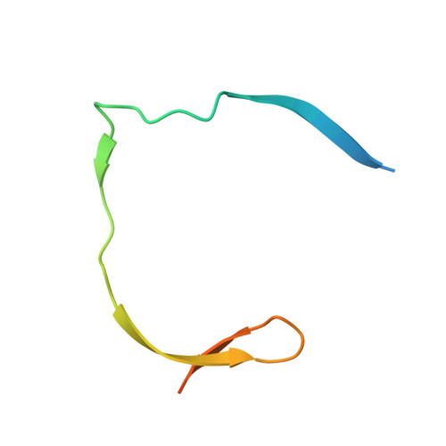
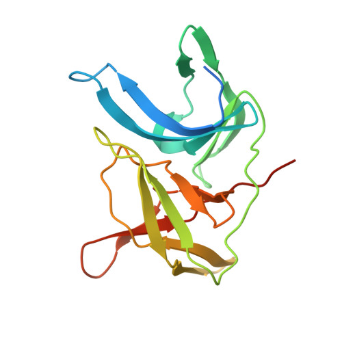
    5H6V

  • PubMed Abstract: 

    The NS2B-NS3 viral protease is an attractive drug target against Zika virus (ZIKV) due to its importance in viral replication and maturation. Here we report the crystal structure of protease in complex with a dipeptide inhibitor, Acyl-KR-aldehyde (compound 1). The aldehyde moiety forms a covalent bond with the catalytic Ser 135 of NS3. The Arg and Lys residues in the inhibitor occupy the S1 and S2 sites of the protease, respectively. Nuclear magnetic resonance studies demonstrate that the complex is in the closed conformation in solution. The chemical environment of residues surrounding the active site is sensitive to the bound inhibitor as demonstrated by the comparison with two other non-covalent dipeptides, Acyl-K-Agmatine (compound 2) and Acyl-KR-COOH (compound 3). Removing the aldehyde moiety in 1 converts the binding mode from a slow to a fast exchange regime. The structural dynamics information obtained in this study will guide future drug discovery against ZIKV and other flaviviruses.


  • Organizational Affiliation

    Experimental Therapeutics Centre, Agency for Science, Technology and Research (A(∗)STAR), 31 Biopolis way, Nanos, #03-01, Singapore 138669, Singapore.


Macromolecules
Find similar proteins by:  (by identity cutoff)  |  3D Structure
Entity ID: 1
MoleculeChains Sequence LengthOrganismDetailsImage
Genome polyprotein52Zika virusMutation(s): 0 
UniProt
Find proteins for H8XX12 (Zika virus)
Explore H8XX12 
Go to UniProtKB:  H8XX12
Entity Groups  
Sequence Clusters30% Identity50% Identity70% Identity90% Identity95% Identity100% Identity
UniProt GroupH8XX12
Sequence Annotations
Expand
  • Reference Sequence
Find similar proteins by:  (by identity cutoff)  |  3D Structure
Entity ID: 2
MoleculeChains Sequence LengthOrganismDetailsImage
Genome polyprotein178Zika virusMutation(s): 1 
UniProt
Find proteins for H8XX12 (Zika virus)
Explore H8XX12 
Go to UniProtKB:  H8XX12
Entity Groups  
Sequence Clusters30% Identity50% Identity70% Identity90% Identity95% Identity100% Identity
UniProt GroupH8XX12
Sequence Annotations
Expand
  • Reference Sequence
Small Molecules
Ligands 1 Unique
IDChains Name / Formula / InChI Key2D Diagram3D Interactions
7HS
Query on 7HS

Download Ideal Coordinates CCD File 
C [auth B](S)-2-acetamido-6-amino-N-((S)-5-guanidino-1-oxopentan-2-yl)hexanamide
C14 H28 N6 O3
VZSVGUHOHBMCOO-RYUDHWBXSA-N
Experimental Data & Validation

Experimental Data

  • Method: X-RAY DIFFRACTION
  • Resolution: 2.42 Å
  • R-Value Free: 0.244 
  • R-Value Work: 0.191 
  • R-Value Observed: 0.194 
  • Space Group: P 43 2 2
Unit Cell:
Length ( Å )Angle ( ˚ )
a = 42.33α = 90
b = 42.33β = 90
c = 214.758γ = 90
Software Package:
Software NamePurpose
PHENIXrefinement
SHELXLdata collection
iMOSFLMdata reduction
Aimlessdata scaling
MOLREPmodel building

Structure Validation

View Full Validation Report


Ligand Structure Quality Assessment 


Entry History & Funding Information

Deposition Data


Funding OrganizationLocationGrant Number
NTUSingaporeSTART UP GRANT
NMRCSingaporeCBRG14May05

Revision History  (Full details and data files)

  • Version 1.0: 2017-06-14
    Type: Initial release
  • Version 1.1: 2018-07-04
    Changes: Data collection, Database references
  • Version 1.2: 2023-11-08
    Changes: Data collection, Database references, Refinement description