Structural basis of autoinhibition and activation of the DNA-targeting ADP-ribosyltransferase pierisin-1
Oda, T., Hirabayashi, H., Shikauchi, G., Takamura, R., Hiraga, K., Minami, H., Hashimoto, H., Yamamoto, M., Wakabayashi, K., Shimizu, T., Sato, M.(2017) J Biol Chem 292: 15445-15455
- PubMed: 28765284
- DOI: https://doi.org/10.1074/jbc.M117.776641
- Primary Citation of Related Structures:
5H6J, 5H6K, 5H6L, 5H6M, 5H6N - PubMed Abstract:
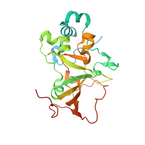
ADP-ribosyltransferases transfer the ADP-ribose moiety of βNAD + to an acceptor molecule, usually a protein that modulates the function of the acceptor. Pierisin-1 is an ADP-ribosyltransferase from the cabbage butterfly Pieris rapae and is composed of N-terminal catalytic and C-terminal ricin B-like domains. Curiously, it ADP-ribosylates the DNA duplex, resulting in apoptosis of various cancer cells, which has raised interest in pierisin-1 as an anti-cancer agent. However, both the structure and the mechanism of DNA ADP-ribosylation are unclear. Here, we report the crystal structures of the N-terminal catalytic domain of pierisin-1, its complex with βNAD + , and the catalytic domain with the linker connecting it to the ricin B-like domains. We found that the catalytic domain possesses a defined, positively charged region on the molecular surface but that its overall structure is otherwise similar to those of protein-targeting ADP-ribosyltransferases. Electrophoretic mobility shift assays and site-directed mutagenesis indicated that pierisin-1 binds double-stranded but not single-stranded DNA and that Lys 122 , Lys 123 , and Lys 124 , which are found in a loop, and Arg 181 and Arg 187 , located in a basic cleft near the loop, are required for DNA binding. Furthermore, the structure of the catalytic domain with the linker revealed an autoinhibitory mechanism in which the linker occupies and blocks both the βNAD + - and DNA-binding sites, suggesting that proteolytic cleavage to remove the linker is necessary for enzyme catalysis. Our study provides a structural basis for the DNA-acceptor specificity of pierisin-1 and reveals that a self-regulatory mechanism is required for its activity.
Organizational Affiliation:
From the Graduate School of Medical Life Science, Yokohama City University, 1-7-29 Suehiro-cho, Tsurumi-ku, Yokohama, Kanagawa 230-0045, Japan.