5H3J

Crystal structure of Grasp domain of Grasp55 complexed with the Golgin45 C-terminus


Experimental Data Snapshot

  • Method: X-RAY DIFFRACTION
  • Resolution: 1.33 Å
  • R-Value Free: 0.162 
  • R-Value Work: 0.137 
  • R-Value Observed: 0.138 

wwPDB Validation   3D Report Full Report


This is version 1.3 of the entry. See complete history


Literature

Structural Basis for the Interaction between Golgi Reassembly-stacking Protein GRASP55 and Golgin45

Zhao, J.Li, B.Huang, X.Morelli, X.Shi, N.

(2017) J Biol Chem 292: 2956-2965

  • DOI: https://doi.org/10.1074/jbc.M116.765990
  • Primary Citation of Related Structures:  
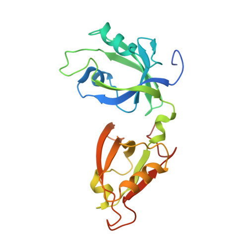
    5H3J

  • PubMed Abstract: 

    Golgin45 is required for normal Golgi structure and the transportation of protein from the ER. It forms a specific complex with GRASP55 in vivo Little is known regarding the molecular details of this interaction and its structural role in stacking of the Golgi complex. Here, we present the crystal structure of the GRASP domains of GRASP55 in complex with the Golgin45 C-terminal peptide, determined at 1.33 Å resolution. Similar to the structure of GRASP65 bound to GM130 reported recently, this structure reveals more than one interacting site and involves both PDZ1 and PDZ2 domains of the GRASP simultaneously. The C-terminal peptides of Golgin45 and GM130 present a conserved PDZ domain binding motif sequence and recognize the canonical PDZ-peptide binding groove of the PDZ1 domains of GRASP55 and GRASP65. A main difference in this recognition process resides in a structural rearrangement of GRASP65-GM130 that does not occur for the GRASP55-Golgin45 complex. The binding site at the cleft between the PDZ1 and PDZ2 domains of GRASP65 is dominated by hydrophobic interactions with GM130 that are not observed in the GRASP55-Golgin45 complex. In addition, a unique zinc finger structure is revealed in the GRASP55-Golgin45 complex crystal structure. Mutagenesis experiments support these structural observations and demonstrate that two of these sites are required to form a stable complex. Finally, a novel Golgi stacking model is proposed according to these structural findings.


  • Organizational Affiliation

    From the State Key Laboratory of Structural Chemistry, Fujian Institute of Research on the Structure of Matter, Chinese Academy of Sciences, Fuzhou 350002, China and.


Macromolecules
Find similar proteins by:  (by identity cutoff)  |  3D Structure
Entity ID: 1
MoleculeChains Sequence LengthOrganismDetailsImage
Golgi reassembly-stacking protein 2211Mus musculusMutation(s): 0 
Gene Names: Gorasp2
UniProt
Find proteins for Q99JX3 (Mus musculus)
Explore Q99JX3 
Go to UniProtKB:  Q99JX3
Entity Groups  
Sequence Clusters30% Identity50% Identity70% Identity90% Identity95% Identity100% Identity
UniProt GroupQ99JX3
Sequence Annotations
Expand
  • Reference Sequence
Find similar proteins by:  (by identity cutoff)  |  3D Structure
Entity ID: 2
MoleculeChains Sequence LengthOrganismDetailsImage
Golgin-4528Mus musculusMutation(s): 0 
Gene Names: Blzf1
UniProt
Find proteins for Q8R2X8 (Mus musculus)
Explore Q8R2X8 
Go to UniProtKB:  Q8R2X8
Entity Groups  
Sequence Clusters30% Identity50% Identity70% Identity90% Identity95% Identity100% Identity
UniProt GroupQ8R2X8
Sequence Annotations
Expand
  • Reference Sequence
Small Molecules
Ligands 1 Unique
IDChains Name / Formula / InChI Key2D Diagram3D Interactions
ZN
Query on ZN

Download Ideal Coordinates CCD File 
C [auth A]ZINC ION
Zn
PTFCDOFLOPIGGS-UHFFFAOYSA-N
Experimental Data & Validation

Experimental Data

  • Method: X-RAY DIFFRACTION
  • Resolution: 1.33 Å
  • R-Value Free: 0.162 
  • R-Value Work: 0.137 
  • R-Value Observed: 0.138 
  • Space Group: C 1 2 1
Unit Cell:
Length ( Å )Angle ( ˚ )
a = 97.681α = 90
b = 35.855β = 93.54
c = 64.03γ = 90
Software Package:
Software NamePurpose
PHENIXrefinement
xia2data reduction
HKL-2000data scaling
PHASERphasing

Structure Validation

View Full Validation Report


Entry History & Funding Information

Deposition Data


Funding OrganizationLocationGrant Number
National Natural Science Foundation of ChinaChina31370738

Revision History  (Full details and data files)

  • Version 1.0: 2017-01-11
    Type: Initial release
  • Version 1.1: 2017-03-01
    Changes: Database references
  • Version 1.2: 2017-10-18
    Changes: Author supporting evidence
  • Version 1.3: 2023-11-08
    Changes: Data collection, Database references, Derived calculations, Refinement description