5H1E

Interaction between vitamin D receptor and coactivator peptide SRC2-3


Experimental Data Snapshot

  • Method: X-RAY DIFFRACTION
  • Resolution: 2.60 Å
  • R-Value Free: 0.248 
  • R-Value Work: 0.239 
  • R-Value Observed: 0.240 

wwPDB Validation   3D Report Full Report


Ligand Structure Quality Assessment 


This is version 1.2 of the entry. See complete history


Literature

SRC2-3 binds to vitamin D receptor with high sensitivity and strong affinity

Egawa, D.Itoh, T.Kato, A.Kataoka, S.Anami, Y.Yamamoto, K.

(2017) Bioorg Med Chem 25: 568-574

  • DOI: https://doi.org/10.1016/j.bmc.2016.11.020
  • Primary Citation of Related Structures:  
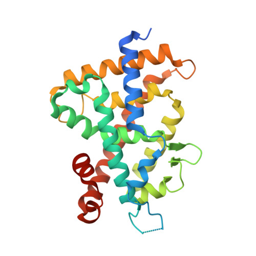
    5H1E

  • PubMed Abstract: 

    Vitamin D receptor (VDR) is a member of the nuclear receptor superfamily and regulates the expression of target genes through ligand binding. To express the target gene, coactivator binding to the VDR/ligand complex is essential. Although there are many coactivators in living cells, precise interactions between coactivators and VDR have not been clarified. Here, we synthesized two coactivator peptides, DRIP205-2 and SRC2-3, evaluated their affinity for the ligand-binding domain (LBD) of VDR using 1α,25-dihydroxyvitamin D 3 , partial agonist 1, and antagonist 2 by surface plasmon resonance (SPR), and assessed their interaction modes with VDR-LBD using X-ray crystallographic analysis. This study showed that the SRC2-3 peptide is more sensitive to the ligands (agonist, partial agonist, and antagonist) and shows more intimate interactions with VDR-LBD than DRIP205-2 peptide.


  • Organizational Affiliation

    Laboratory of Drug Design and Medicinal Chemistry, Showa Pharmaceutical University, 3-3165 Higashi-Tamagawagakuen, Machida, Tokyo 194-8543, Japan.


Macromolecules
Find similar proteins by:  (by identity cutoff)  |  3D Structure
Entity ID: 1
MoleculeChains Sequence LengthOrganismDetailsImage
Vitamin D3 receptor271Rattus norvegicusMutation(s): 0 
Gene Names: VdrNr1i1
UniProt
Find proteins for P13053 (Rattus norvegicus)
Explore P13053 
Go to UniProtKB:  P13053
Entity Groups  
Sequence Clusters30% Identity50% Identity70% Identity90% Identity95% Identity100% Identity
UniProt GroupP13053
Sequence Annotations
Expand
  • Reference Sequence

Find similar proteins by:  Sequence   |   3D Structure  

Entity ID: 2
MoleculeChains Sequence LengthOrganismDetailsImage
Nuclear receptor coactivator 2 peptideB [auth C]13Homo sapiensMutation(s): 0 
UniProt & NIH Common Fund Data Resources
Find proteins for Q15596 (Homo sapiens)
Explore Q15596 
Go to UniProtKB:  Q15596
PHAROS:  Q15596
GTEx:  ENSG00000140396 
Entity Groups  
Sequence Clusters30% Identity50% Identity70% Identity90% Identity95% Identity100% Identity
UniProt GroupQ15596
Sequence Annotations
Expand
  • Reference Sequence
Small Molecules
Ligands 1 Unique
IDChains Name / Formula / InChI Key2D Diagram3D Interactions
VDX
Query on VDX

Download Ideal Coordinates CCD File 
C [auth A]5-{2-[1-(5-HYDROXY-1,5-DIMETHYL-HEXYL)-7A-METHYL-OCTAHYDRO-INDEN-4-YLIDENE]-ETHYLIDENE}-4-METHYLENE-CYCLOHEXANE-1,3-DIOL
C27 H44 O3
GMRQFYUYWCNGIN-NKMMMXOESA-N
Binding Affinity Annotations 
IDSourceBinding Affinity
VDX BindingDB:  5H1E Ki: min: 0.04, max: 1 (nM) from 5 assay(s)
EC50: min: 0.05, max: 0.3 (nM) from 4 assay(s)
Experimental Data & Validation

Experimental Data

  • Method: X-RAY DIFFRACTION
  • Resolution: 2.60 Å
  • R-Value Free: 0.248 
  • R-Value Work: 0.239 
  • R-Value Observed: 0.240 
  • Space Group: C 1 2 1
Unit Cell:
Length ( Å )Angle ( ˚ )
a = 155.77α = 90
b = 42.88β = 95.83
c = 42.21γ = 90
Software Package:
Software NamePurpose
REFMACrefinement

Structure Validation

View Full Validation Report


Ligand Structure Quality Assessment 


Entry History 

Deposition Data

Revision History  (Full details and data files)

  • Version 1.0: 2017-01-11
    Type: Initial release
  • Version 1.1: 2020-02-26
    Changes: Data collection
  • Version 1.2: 2024-03-20
    Changes: Data collection, Database references