5H06

Crystal structure of AmyP in complex with maltose


Experimental Data Snapshot

  • Method: X-RAY DIFFRACTION
  • Resolution: 1.95 Å
  • R-Value Free: 0.212 
  • R-Value Work: 0.164 
  • R-Value Observed: 0.166 

wwPDB Validation   3D Report Full Report


This is version 2.1 of the entry. See complete history


Literature

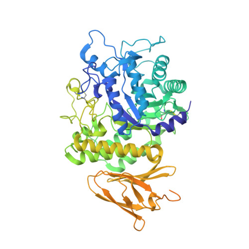
Crystal structure of a raw-starch-degrading bacterial alpha-amylase belonging to subfamily 37 of the glycoside hydrolase family GH13

Liu, Y.Yu, J.Li, F.Peng, H.Zhang, X.Xiao, Y.He, C.

(2017) Sci Rep 7: 44067-44067

  • DOI: https://doi.org/10.1038/srep44067
  • Primary Citation of Related Structures:  
    5H05, 5H06

  • PubMed Abstract: 

    Subfamily 37 of the glycoside hydrolase family GH13 was recently established on the basis of the discovery of a novel α-amylase, designated AmyP, from a marine metagenomic library. AmyP exhibits raw-starch-degrading activity and consists of an N-terminal catalytic domain and a C-terminal starch-binding domain. To understand this newest subfamily, we determined the crystal structure of the catalytic domain of AmyP, named AmyP ΔSBD , complexed with maltose, and the crystal structure of the E221Q mutant AmyP ΔSBD complexed with maltotriose. Glu221 is one of the three conserved catalytic residues, and AmyP is inactivated by the E221Q mutation. Domain B of AmyP ΔSBD forms a loop that protrudes from domain A, stabilizes the conformation of the active site and increases the thermostability of the enzyme. A new calcium ion is situated adjacent to the -3 subsite binding loop and may be responsible for the increased thermostability of the enzyme after the addition of calcium. Moreover, Tyr36 participates in both stacking and hydrogen bonding interactions with the sugar motif at subsite -3. This work provides the first insights into the structure of α-amylases belonging to subfamily 37 of GH13 and may contribute to the rational design of α-amylase mutants with enhanced performance in biotechnological applications.


  • Organizational Affiliation

    Anhui Provincial Engineering Technology Research Center of Microorganisms and Biocatalysis and School of Life Sciences, Anhui University, Hefei, Anhui 230601, China.


Macromolecules
Find similar proteins by:  (by identity cutoff)  |  3D Structure
Entity ID: 1
MoleculeChains Sequence LengthOrganismDetailsImage
AmyP
A, B, C, D
640marine metagenomeMutation(s): 0 
Entity Groups  
Sequence Clusters30% Identity50% Identity70% Identity90% Identity95% Identity100% Identity
Sequence Annotations
Expand
  • Reference Sequence
Oligosaccharides

Help

Entity ID: 2
MoleculeChains Length2D Diagram Glycosylation3D Interactions
alpha-D-glucopyranose-(1-4)-alpha-D-glucopyranose
E, F, G, H
2N/A
Glycosylation Resources
GlyTouCan:  G07411ON
GlyCosmos:  G07411ON
Experimental Data & Validation

Experimental Data

  • Method: X-RAY DIFFRACTION
  • Resolution: 1.95 Å
  • R-Value Free: 0.212 
  • R-Value Work: 0.164 
  • R-Value Observed: 0.166 
  • Space Group: P 21 21 21
Unit Cell:
Length ( Å )Angle ( ˚ )
a = 79.87α = 90
b = 130.532β = 90
c = 217.53γ = 90
Software Package:
Software NamePurpose
PHENIXrefinement
HKL-2000data collection
HKL-2000data scaling
PDB_EXTRACTdata extraction
HKL-2000data reduction
PHASERphasing

Structure Validation

View Full Validation Report


Entry History 

Deposition Data

  • Released Date: 2017-08-16 
  • Deposition Author(s): He, C., Liu, Y.

Revision History  (Full details and data files)

  • Version 1.0: 2017-08-16
    Type: Initial release
  • Version 2.0: 2020-07-29
    Type: Remediation
    Reason: Carbohydrate remediation
    Changes: Atomic model, Data collection, Derived calculations, Non-polymer description, Structure summary
  • Version 2.1: 2023-11-08
    Changes: Data collection, Database references, Refinement description, Structure summary