Structural studies of the periplasmic portion of the diguanylate cyclase CdgH from Vibrio cholerae.
Xu, M., Wang, Y.Z., Yang, X.A., Jiang, T., Xie, W.(2017) Sci Rep 7: 1861-1861
- PubMed: 28500346
- DOI: https://doi.org/10.1038/s41598-017-01989-6
- Primary Citation of Related Structures:
5GZS - PubMed Abstract:
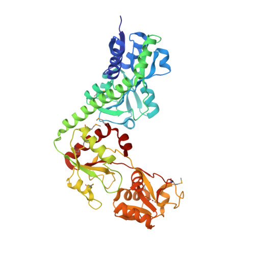
Cyclic diguanylate monophosphate (c-di-GMP) is a second messenger involved in bacterial signal transduction and produced by diguanylate cyclases (DGCs) generally containing highly variable periplasmic signal-recognition domains. CdgH is a DGC enzyme that regulates rugosity associated phenotypes in Vibrio cholerae. CdgH has two N-terminal tandem periplasmic substrate-binding (PBPb) domains for its signal recognition; however, the role of the tandem PBPb domains remains unclear. Here, we reported the crystal structure of the periplasmic portion of CdgH, which indicated that both tandem PBPb domains consist of typical interlobe ligand-binding architecture. Unexpectedly, the PBPb-I domain binds an L-arginine which apparently has been co-purified from the E. coli expression system, whereas the PBPb-II domain is in an unliganded open state. Structural comparison with other amino acid-binding proteins indicated that despite similar ligand-binding pockets, the PBPb-I domain possesses two ligand-binding residues (E122 and Y148) not conserved in homologs and involved in hydrophilic and hydrophobic interactions with L-arginine. Isothermal titration calorimetry indicated that the PBPb-I is primarily an L-arginine/L-lysine/L-ornithine-binding domain, whereas the PBPb-II domain exhibits a preference for L-glutamine and L-histidine. Remarkably, we found that the periplasmic portion of CdgH forms a stable dimer in solution and L-arginine binding would cause conformational changes of the dimer.
Organizational Affiliation:
National Laboratory of Biomacromolecules, CAS Center for Excellence in Biomacromolecules, Institute of Biophysics, Chinese Academy of Sciences, Beijing, China. xumin@moon.ibp.ac.cn.