


Cyclodeaminase_PA
Ying, H., Chen, K.To be published.
Experimental Data Snapshot
Entity ID: 1 | |||||
|---|---|---|---|---|---|
| Molecule | Chains | Sequence Length | Organism | Details | Image |
| Lysine cyclodeaminase | A [auth B], B [auth A] | 359 | Streptomyces pristinaespiralis | Mutation(s): 0 Gene Names: pipA, SPRI_0308, SPRI_7045 EC: 4.3.1.12 | 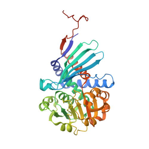 |
UniProt | |||||
Find proteins for D9UBW0 (Streptomyces pristinaespiralis) Explore D9UBW0 Go to UniProtKB: D9UBW0 | |||||
Entity Groups | |||||
| Sequence Clusters | 30% Identity50% Identity70% Identity90% Identity95% Identity100% Identity | ||||
| UniProt Group | D9UBW0 | ||||
Sequence AnnotationsExpand | |||||
| |||||
| Ligands 2 Unique | |||||
|---|---|---|---|---|---|
| ID | Chains | Name / Formula / InChI Key | 2D Diagram | 3D Interactions | |
| NAD Query on NAD | C [auth B], E [auth A] | NICOTINAMIDE-ADENINE-DINUCLEOTIDE C21 H27 N7 O14 P2 BAWFJGJZGIEFAR-NNYOXOHSSA-N |  | ||
| YCP Query on YCP | D [auth B], F [auth A] | (2S)-piperidine-2-carboxylic acid C6 H11 N O2 HXEACLLIILLPRG-YFKPBYRVSA-N |  | ||
| Length ( Å ) | Angle ( ˚ ) |
|---|---|
| a = 136.926 | α = 90 |
| b = 74.06 | β = 132.72 |
| c = 95.718 | γ = 90 |
| Software Name | Purpose |
|---|---|
| REFMAC | refinement |
| HKL-3000 | data reduction |
| HKL-3000 | data scaling |
| Funding Organization | Location | Grant Number |
|---|---|---|
| China | -- |
RCSB PDB (citation) is hosted by
RCSB PDB is a member of the

