5GYK

Crystal Structure of Mdm12-deletion mutant


Experimental Data Snapshot

  • Method: X-RAY DIFFRACTION
  • Resolution: 3.60 Å
  • R-Value Free: 0.267 
  • R-Value Work: 0.220 
  • R-Value Observed: 0.222 

wwPDB Validation   3D Report Full Report


Ligand Structure Quality Assessment 


This is version 2.0 of the entry. See complete history


Literature

Crystal structure of Mdm12 reveals the architecture and dynamic organization of the ERMES complex

Jeong, H.Park, J.Lee, C.

(2016) EMBO Rep 17: 1857-1871

  • DOI: https://doi.org/10.15252/embr.201642706
  • Primary Citation of Related Structures:  
    5GYD, 5GYK

  • PubMed Abstract: 

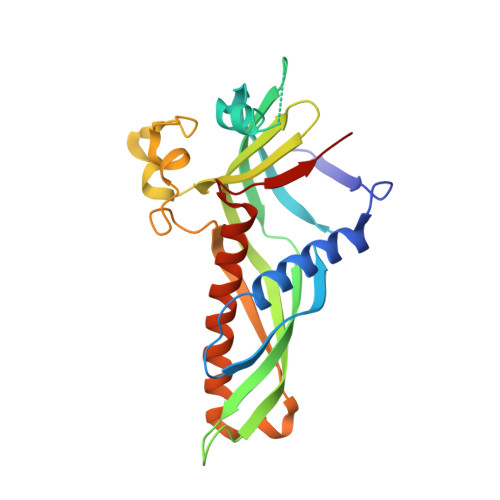
    The endoplasmic reticulum-mitochondria encounter structure (ERMES) is a protein complex that plays a tethering role in physically connecting ER and mitochondria membranes. The ERMES complex is composed of Mdm12, Mmm1, and Mdm34, which have a SMP domain in common, and Mdm10. Here, we report the crystal structure of S. cerevisiae Mdm12. The Mdm12 forms a dimeric SMP structure through domain swapping of the β1-strand comprising residues 1-7. Biochemical experiments reveal a phospholipid-binding site located along a hydrophobic channel of the Mdm12 structure and that Mdm12 might have a binding preference for glycerophospholipids harboring a positively charged head group. Strikingly, both full-length Mdm12 and Mdm12 truncated to exclude the disordered region (residues 74-114) display the same organization in the asymmetric unit, although they crystallize as a tetramer and hexamer, respectively. Taken together, these studies provide a novel understanding of the overall organization of SMP domains in the ERMES complex, indicating that Mdm12 interacts with Mdm34 through head-to-head contact, and with Mmm1 through tail-to-tail contact of SMP domains.


  • Organizational Affiliation

    Department of Biological Sciences, School of Life Sciences Ulsan National Institute of Science and Technology, Ulsan, Korea.


Macromolecules
Find similar proteins by:  (by identity cutoff)  |  3D Structure
Entity ID: 1
MoleculeChains Sequence LengthOrganismDetailsImage
Mitochondrial distribution and morphology protein 12
A, B, C, D, E
A, B, C, D, E, F
236Saccharomyces cerevisiae S288CMutation(s): 0 
Gene Names: MDM12YOL009C
UniProt
Find proteins for Q92328 (Saccharomyces cerevisiae (strain ATCC 204508 / S288c))
Explore Q92328 
Go to UniProtKB:  Q92328
Entity Groups  
Sequence Clusters30% Identity50% Identity70% Identity90% Identity95% Identity100% Identity
UniProt GroupQ92328
Sequence Annotations
Expand
  • Reference Sequence
Small Molecules
Ligands 1 Unique
IDChains Name / Formula / InChI Key2D Diagram3D Interactions
PEE
Query on PEE

Download Ideal Coordinates CCD File 
G [auth A],
H [auth D]
1,2-dioleoyl-sn-glycero-3-phosphoethanolamine
C41 H78 N O8 P
MWRBNPKJOOWZPW-NYVOMTAGSA-N
Experimental Data & Validation

Experimental Data

  • Method: X-RAY DIFFRACTION
  • Resolution: 3.60 Å
  • R-Value Free: 0.267 
  • R-Value Work: 0.220 
  • R-Value Observed: 0.222 
  • Space Group: P 21 21 21
Unit Cell:
Length ( Å )Angle ( ˚ )
a = 109.239α = 90
b = 148.243β = 90
c = 212.394γ = 90
Software Package:
Software NamePurpose
PHENIXrefinement
HKL-2000data processing
AutoSolphasing
Cootmodel building

Structure Validation

View Full Validation Report


Ligand Structure Quality Assessment 


Entry History 

Deposition Data

Revision History  (Full details and data files)

  • Version 1.0: 2016-11-16
    Type: Initial release
  • Version 1.1: 2016-12-14
    Changes: Database references
  • Version 2.0: 2023-11-08
    Changes: Advisory, Atomic model, Data collection, Database references, Derived calculations, Non-polymer description, Refinement description, Structure summary