5GMV

LC3B-FUNDC1 complex


Experimental Data Snapshot

  • Method: X-RAY DIFFRACTION
  • Resolution: 2.25 Å
  • R-Value Free: 0.272 
  • R-Value Work: 0.206 
  • R-Value Observed: 0.209 

wwPDB Validation   3D Report Full Report


This is version 1.1 of the entry. See complete history


Literature

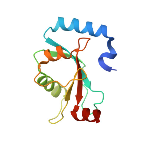
Structural insights into the recognition of phosphorylated FUNDC1 by LC3B in mitophagy

Lv, M.Wang, C.Li, F.Peng, J.Wen, B.Gong, Q.Shi, Y.Tang, Y.

(2017) Protein Cell 8: 25-38

  • DOI: https://doi.org/10.1007/s13238-016-0328-8
  • Primary Citation of Related Structures:  
    5GMV

  • PubMed Abstract: 

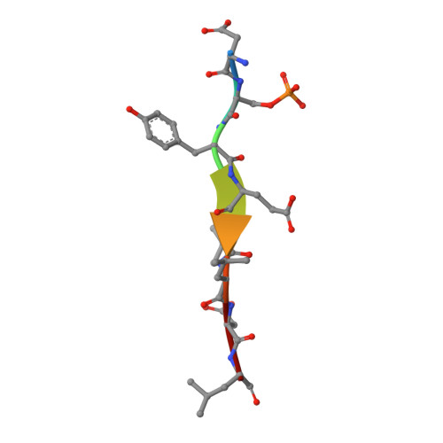
    Mitophagy is an essential intracellular process that eliminates dysfunctional mitochondria and maintains cellular homeostasis. Mitophagy is regulated by the post-translational modification of mitophagy receptors. Fun14 domain-containing protein 1 (FUNDC1) was reported to be a new receptor for hypoxia-induced mitophagy in mammalian cells and interact with microtubule-associated protein light chain 3 beta (LC3B) through its LC3 interaction region (LIR). Moreover, the phosphorylation modification of FUNDC1 affects its binding affinity for LC3B and regulates selective mitophagy. However, the structural basis of this regulation mechanism remains unclear. Here, we present the crystal structure of LC3B in complex with a FUNDC1 LIR peptide phosphorylated at Ser17 (pS 17 ), demonstrating the key residues of LC3B for the specific recognition of the phosphorylated or dephosphorylated FUNDC1. Intriguingly, the side chain of LC3B Lys49 shifts remarkably and forms a hydrogen bond and electrostatic interaction with the phosphate group of FUNDC1 pS 17 . Alternatively, phosphorylated Tyr18 (pY 18 ) and Ser13 (pS 13 ) in FUNDC1 significantly obstruct their interaction with the hydrophobic pocket and Arg10 of LC3B, respectively. Structural observations are further validated by mutation and isothermal titration calorimetry (ITC) assays. Therefore, our structural and biochemical results reveal a working model for the specific recognition of FUNDC1 by LC3B and imply that the reversible phosphorylation modification of mitophagy receptors may be a switch for selective mitophagy.


  • Organizational Affiliation

    Hefei National Laboratory for Physical Sciences at Microscale and School of Life Sciences, University of Science and Technology of China, Hefei, 230027, China.


Macromolecules
Find similar proteins by:  (by identity cutoff)  |  3D Structure
Entity ID: 1
MoleculeChains Sequence LengthOrganismDetailsImage
Microtubule-associated proteins 1A/1B light chain 3BA [auth B],
C [auth A]
125Homo sapiensMutation(s): 0 
Gene Names: MAP1LC3BMAP1ALC3
UniProt & NIH Common Fund Data Resources
Find proteins for Q9GZQ8 (Homo sapiens)
Explore Q9GZQ8 
Go to UniProtKB:  Q9GZQ8
PHAROS:  Q9GZQ8
GTEx:  ENSG00000140941 
Entity Groups  
Sequence Clusters30% Identity50% Identity70% Identity90% Identity95% Identity100% Identity
UniProt GroupQ9GZQ8
Sequence Annotations
Expand
  • Reference Sequence

Find similar proteins by:  Sequence   |   3D Structure  

Entity ID: 2
MoleculeChains Sequence LengthOrganismDetailsImage
Peptide from FUN14 domain-containing protein 1B [auth D],
D [auth C]
8Homo sapiensMutation(s): 0 
UniProt & NIH Common Fund Data Resources
Find proteins for Q8IVP5 (Homo sapiens)
Explore Q8IVP5 
Go to UniProtKB:  Q8IVP5
PHAROS:  Q8IVP5
GTEx:  ENSG00000069509 
Entity Groups  
UniProt GroupQ8IVP5
Sequence Annotations
Expand
  • Reference Sequence
Small Molecules
Modified Residues  1 Unique
IDChains TypeFormula2D DiagramParent
SEP
Query on SEP
B [auth D],
D [auth C]
L-PEPTIDE LINKINGC3 H8 N O6 PSER
Experimental Data & Validation

Experimental Data

  • Method: X-RAY DIFFRACTION
  • Resolution: 2.25 Å
  • R-Value Free: 0.272 
  • R-Value Work: 0.206 
  • R-Value Observed: 0.209 
  • Space Group: P 1 21 1
Unit Cell:
Length ( Å )Angle ( ˚ )
a = 40.537α = 90
b = 86.85β = 110.86
c = 40.537γ = 90
Software Package:
Software NamePurpose
SCALAdata scaling
REFMACrefinement
PDB_EXTRACTdata extraction
MOSFLMdata reduction
PHASERphasing

Structure Validation

View Full Validation Report


Entry History 

Deposition Data

Revision History  (Full details and data files)

  • Version 1.0: 2017-03-08
    Type: Initial release
  • Version 1.1: 2023-11-08
    Changes: Data collection, Database references, Refinement description