5GIE

Crystal structure of VDR in complex with DLAM-4P (P21 form)


Experimental Data Snapshot

  • Method: X-RAY DIFFRACTION
  • Resolution: 2.39 Å
  • R-Value Free: 0.234 
  • R-Value Work: 0.207 
  • R-Value Observed: 0.209 

wwPDB Validation   3D Report Full Report


Ligand Structure Quality Assessment 


This is version 1.2 of the entry. See complete history


Literature

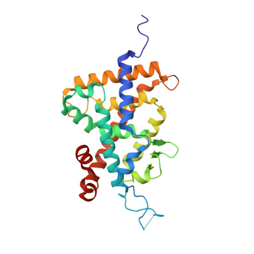
Regulation of the vitamin D receptor by vitamin D lactam derivatives.

Asano, L.Waku, T.Abe, R.Kuwabara, N.Ito, I.Yanagisawa, J.Nagasawa, K.Shimizu, T.

(2016) FEBS Lett 590: 3270-3279

  • DOI: https://doi.org/10.1002/1873-3468.12348
  • Primary Citation of Related Structures:  
    5GIC, 5GID, 5GIE

  • PubMed Abstract: 

    The active metabolite of vitamin D3 , 1α,25-dihydroxyvitamin D3 , acts as a ligand for the vitamin D receptor (VDR) and activates VDR-mediated gene expression. Recently, we characterized 1α,25-dihydroxyvitamin D3 -26,23-lactams (DLAMs), which mimic vitamin D3 metabolites, as noncalcemic VDR ligands that barely activate the receptor. In this study, we present structural insights onto the regulation of VDR function by DLAMs. X-ray crystallographic analysis revealed that DLAMs induced a large conformational change in the loop region between helices H6 and H7 in the VDR ligand-binding domain. Our structural analysis suggests that targeting of the loop region may be a new mode of VDR regulation.


  • Organizational Affiliation

    Institute for Chemical Research, Kyoto University, Uji, Kyoto, Japan.


Macromolecules
Find similar proteins by:  (by identity cutoff)  |  3D Structure
Entity ID: 1
MoleculeChains Sequence LengthOrganismDetailsImage
Vitamin D3 receptorA,
C [auth D]
256Rattus norvegicusMutation(s): 0 
Gene Names: VdrNr1i1
UniProt
Find proteins for P13053 (Rattus norvegicus)
Explore P13053 
Go to UniProtKB:  P13053
Entity Groups  
Sequence Clusters30% Identity50% Identity70% Identity90% Identity95% Identity100% Identity
UniProt GroupP13053
Sequence Annotations
Expand
  • Reference Sequence

Find similar proteins by:  Sequence   |   3D Structure  

Entity ID: 2
MoleculeChains Sequence LengthOrganismDetailsImage
SRC1B [auth C],
D [auth E]
10Homo sapiensMutation(s): 0 
UniProt & NIH Common Fund Data Resources
Find proteins for Q15648 (Homo sapiens)
Explore Q15648 
Go to UniProtKB:  Q15648
PHAROS:  Q15648
GTEx:  ENSG00000125686 
Entity Groups  
Sequence Clusters30% Identity50% Identity70% Identity90% Identity95% Identity100% Identity
UniProt GroupQ15648
Sequence Annotations
Expand
  • Reference Sequence
Small Molecules
Ligands 1 Unique
IDChains Name / Formula / InChI Key2D Diagram3D Interactions
VDP
Query on VDP

Download Ideal Coordinates CCD File 
E [auth A],
F [auth D]
(3R,5S)-5-[(2R)-2-[(1R,3aS,4E,7aR)-7a-methyl-4-[(2Z)-2-[(3S,5R)-2-methylidene-3,5-bis(oxidanyl)cyclohexylidene]ethylidene]-2,3,3a,5,6,7-hexahydro-1H-inden-1-yl]propyl]-3-methyl-3-oxidanyl-1-(4-phenylbutyl)pyrrolidin-2-one
C37 H53 N O4
IVUJLUOFPHOBPG-HEMHZJQPSA-N
Binding Affinity Annotations 
IDSourceBinding Affinity
VDP Binding MOAD:  5GIE Kd: 769.2 (nM) from 1 assay(s)
Experimental Data & Validation

Experimental Data

  • Method: X-RAY DIFFRACTION
  • Resolution: 2.39 Å
  • R-Value Free: 0.234 
  • R-Value Work: 0.207 
  • R-Value Observed: 0.209 
  • Space Group: P 1 21 1
Unit Cell:
Length ( Å )Angle ( ˚ )
a = 65.364α = 90
b = 42.077β = 94.55
c = 96.802γ = 90
Software Package:
Software NamePurpose
PHENIXrefinement
XDSdata reduction
XDSdata scaling
MOLREPphasing

Structure Validation

View Full Validation Report


Ligand Structure Quality Assessment 


Entry History 

Deposition Data

Revision History  (Full details and data files)

  • Version 1.0: 2016-12-07
    Type: Initial release
  • Version 1.1: 2019-08-28
    Changes: Data collection, Database references, Derived calculations
  • Version 1.2: 2024-03-20
    Changes: Data collection, Database references