


5G6U
Crystal structure of langerin carbohydrate recognition domain with GlcNS6S
- PDB DOI: https://doi.org/10.2210/pdb5G6U/pdb
- Classification: CARBOHYDRATE BINDING PROTEIN
- Organism(s): Homo sapiens
- Expression System: Escherichia coli BL21(DE3)
- Mutation(s): No
- Deposited: 2016-07-21 Released: 2018-02-21
Experimental Data Snapshot
- Method: X-RAY DIFFRACTION
- Resolution: 1.84 Å
- R-Value Free: 0.180
- R-Value Work: 0.152
- R-Value Observed: 0.153

This is version 2.1 of the entry. See complete history.

Macromolecules
Find similar proteins by:
(by identity cutoff) | 3D Structure
Entity ID: 1 | |||||
|---|---|---|---|---|---|
| Molecule | Chains | Sequence Length | Organism | Details | Image |
| LANGERIN | 261 | Homo sapiens | Mutation(s): 0 | 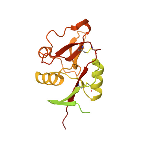 | |
UniProt & NIH Common Fund Data Resources | |||||
Find proteins for Q9UJ71 (Homo sapiens) Explore Q9UJ71 Go to UniProtKB: Q9UJ71 | |||||
PHAROS: Q9UJ71 GTEx: ENSG00000116031 | |||||
Entity Groups | |||||
| Sequence Clusters | 30% Identity50% Identity70% Identity90% Identity95% Identity100% Identity | ||||
| UniProt Group | Q9UJ71 | ||||
Sequence AnnotationsExpand | |||||
| |||||
Small Molecules
| Ligands 5 Unique | |||||
|---|---|---|---|---|---|
| ID | Chains | Name / Formula / InChI Key | 2D Diagram | 3D Interactions | |
| SGN Query on SGN | CA [auth D], F [auth A], L [auth B], U [auth C] | 2-deoxy-6-O-sulfo-2-(sulfoamino)-alpha-D-glucopyranose C6 H13 N O11 S2 DQTRACMFIGDHSN-UKFBFLRUSA-N |  | ||
| TRP Query on TRP | BA [auth D], E [auth A], K [auth B] | TRYPTOPHAN C11 H12 N2 O2 QIVBCDIJIAJPQS-VIFPVBQESA-N |  | ||
| EU Query on EU | G [auth A] J [auth A] M [auth B] T [auth B] W [auth C] | EUROPIUM ION Eu MGVUQZZTJGLWJV-UHFFFAOYSA-N |  | ||
| CA Query on CA | DA [auth D], H [auth A], N [auth B], V [auth C] | CALCIUM ION Ca BHPQYMZQTOCNFJ-UHFFFAOYSA-N |  | ||
| CL Query on CL | AA [auth C] EA [auth D] FA [auth D] GA [auth D] I [auth A] | CHLORIDE ION Cl VEXZGXHMUGYJMC-UHFFFAOYSA-M |  | ||
Experimental Data & Validation
Experimental Data
- Method: X-RAY DIFFRACTION
- Resolution: 1.84 Å
- R-Value Free: 0.180
- R-Value Work: 0.152
- R-Value Observed: 0.153
- Space Group: P 42
Unit Cell:
| Length ( Å ) | Angle ( ˚ ) |
|---|---|
| a = 79.483 | α = 90 |
| b = 79.483 | β = 90 |
| c = 90.71 | γ = 90 |
| Software Name | Purpose |
|---|---|
| PHENIX | refinement |
| XDS | data reduction |
| SCALA | data scaling |
| PHASER | phasing |
Entry History
Deposition Data
- Released Date: 2018-02-21 Deposition Author(s): Porkolab, V., Chabrol, E., Varga, N., Ordanini, S., Sutkeviciute, I., Thepaut, M., Bernardi, A., Fieschi, F.
Revision History (Full details and data files)
- Version 1.0: 2018-02-21
Type: Initial release - Version 1.1: 2018-02-28
Changes: Derived calculations - Version 1.2: 2018-03-28
Changes: Database references - Version 2.0: 2020-07-29
Type: Remediation
Reason: Carbohydrate remediation
Changes: Advisory, Atomic model, Data collection, Derived calculations, Non-polymer description, Structure summary - Version 2.1: 2024-01-10
Changes: Data collection, Database references, Refinement description
