5G15

Structure Aurora A (122-403) bound to activating monobody Mb1 and AMPPCP


Experimental Data Snapshot

  • Method: X-RAY DIFFRACTION
  • Resolution: 2.06 Å
  • R-Value Free: 0.274 
  • R-Value Work: 0.227 
  • R-Value Observed: 0.229 

wwPDB Validation   3D Report Full Report


Ligand Structure Quality Assessment 


This is version 1.3 of the entry. See complete history


Literature

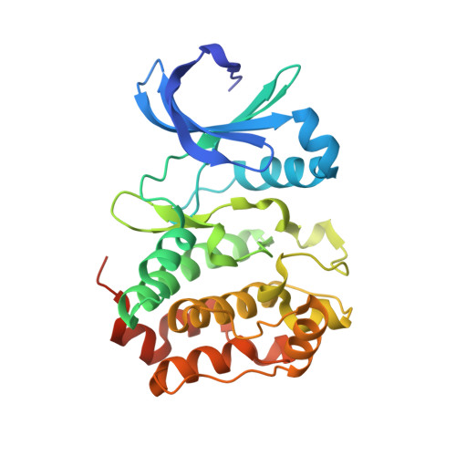
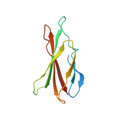
Allosteric modulation of a human protein kinase with monobodies.

Zorba, A.Nguyen, V.Koide, A.Hoemberger, M.Zheng, Y.Kutter, S.Kim, C.Koide, S.Kern, D.

(2019) Proc Natl Acad Sci U S A 116: 13937-13942

  • DOI: https://doi.org/10.1073/pnas.1906024116
  • Primary Citation of Related Structures:  
    5G15, 6C83

  • PubMed Abstract: 

    Despite being the subject of intense effort and scrutiny, kinases have proven to be consistently challenging targets in inhibitor drug design. A key obstacle has been promiscuity and consequent adverse effects of drugs targeting the ATP binding site. Here we introduce an approach to controlling kinase activity by using monobodies that bind to the highly specific regulatory allosteric pocket of the oncoprotein Aurora A (AurA) kinase, thereby offering the potential for more specific kinase modulators. Strikingly, we identify a series of highly specific monobodies acting either as strong kinase inhibitors or activators via differential recognition of structural motifs in the allosteric pocket. X-ray crystal structures comparing AurA bound to activating vs inhibiting monobodies reveal the atomistic mechanism underlying allosteric modulation. The results reveal 3 major advantages of targeting allosteric vs orthosteric sites: extreme selectivity, ability to inhibit as well as activate, and avoidance of competing with ATP that is present at high concentrations in the cells. We envision that exploiting allosteric networks for inhibition or activation will provide a general, powerful pathway toward rational drug design.


  • Organizational Affiliation

    Department of Biochemistry, Brandeis University, Waltham, MA 02454.


Macromolecules
Find similar proteins by:  (by identity cutoff)  |  3D Structure
Entity ID: 1
MoleculeChains Sequence LengthOrganismDetailsImage
AURORA A KINASE282Homo sapiensMutation(s): 0 
EC: 2.7.11.1
UniProt & NIH Common Fund Data Resources
Find proteins for O14965 (Homo sapiens)
Explore O14965 
Go to UniProtKB:  O14965
PHAROS:  O14965
GTEx:  ENSG00000087586 
Entity Groups  
Sequence Clusters30% Identity50% Identity70% Identity90% Identity95% Identity100% Identity
UniProt GroupO14965
Sequence Annotations
Expand
  • Reference Sequence
Find similar proteins by:  (by identity cutoff)  |  3D Structure
Entity ID: 2
MoleculeChains Sequence LengthOrganismDetailsImage
MB1 MONOBODY96synthetic constructMutation(s): 0 
Entity Groups  
Sequence Clusters30% Identity50% Identity70% Identity90% Identity95% Identity100% Identity
Sequence Annotations
Expand
  • Reference Sequence
Experimental Data & Validation

Experimental Data

  • Method: X-RAY DIFFRACTION
  • Resolution: 2.06 Å
  • R-Value Free: 0.274 
  • R-Value Work: 0.227 
  • R-Value Observed: 0.229 
  • Space Group: I 2 2 2
Unit Cell:
Length ( Å )Angle ( ˚ )
a = 75.339α = 90
b = 91.361β = 90
c = 143.715γ = 90
Software Package:
Software NamePurpose
DIALSdata reduction
Aimlessdata scaling
MOLREPphasing
PHASERphasing
REFMACrefinement

Structure Validation

View Full Validation Report


Ligand Structure Quality Assessment 


Entry History 

Deposition Data

Revision History  (Full details and data files)

  • Version 1.0: 2018-03-14
    Type: Initial release
  • Version 1.1: 2019-05-08
    Changes: Data collection, Experimental preparation
  • Version 1.2: 2019-07-24
    Changes: Data collection, Database references, Structure summary
  • Version 1.3: 2024-01-10
    Changes: Data collection, Database references, Derived calculations, Refinement description