5FX8

Complete structure of manganese lipoxygenase of Gaeumannomyces graminis and partial structure of zonadhesin of Komagataella pastoris


Experimental Data Snapshot

  • Method: X-RAY DIFFRACTION
  • Resolution: 2.60 Å
  • R-Value Free: 0.245 
  • R-Value Work: 0.195 
  • R-Value Observed: 0.198 

wwPDB Validation   3D Report Full Report


Ligand Structure Quality Assessment 


This is version 2.1 of the entry. See complete history


Literature

Crystal Structure of Linoleate 13R-Manganese Lipoxygenase and an Adhesion Protein

Chen, Y.Wennman, A.Karkehabadi, S.Engstrom, A.Oliw, E.H.

(2016) J Lipid Res 57: 1574

  • DOI: https://doi.org/10.1194/jlr.M069617
  • Primary Citation of Related Structures:  
    5FX8

  • PubMed Abstract: 

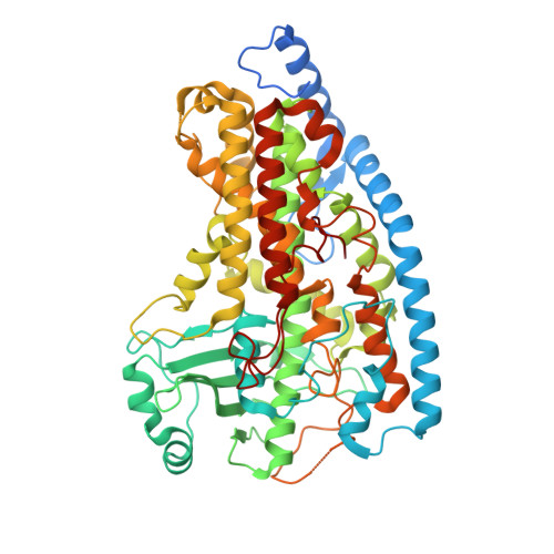
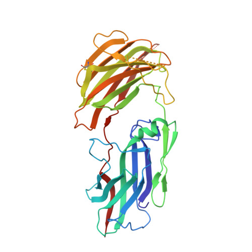
    The crystal structure of 13R-manganese lipoxygenase (MnLOX) of Gaeumannomyces graminis (Gg) in complex with zonadhesin of Pichia pastoris was solved by molecular replacement. Zonadhesin contains β-strands in two subdomains. A comparison of Gg-MnLOX with the 9S-MnLOX of Magnaporthe oryzae (Mo) shows that the protein fold and the geometry of the metal ligands are conserved. The U-shaped active sites differ mainly due to hydrophobic residues of the substrate channel. The volumes and two hydrophobic side pockets near the catalytic base may sanction oxygenation at C-13 and C-9, respectively. Gly-332 of Gg-MnLOX is positioned in the substrate channel between the entrance and the metal center. Replacements with larger residues could restrict oxygen and substrate to reach the active site. C18 fatty acids are likely positioned with C-11 between Mn(2+)OH2 and Leu-336 for hydrogen abstraction and with one side of the 12Z double bond shielded by Phe-337 to prevent antarafacial oxygenation at C-13 and C-11. Phe-347 is positioned at the end of the substrate channel and replacement with smaller residues can position C18 fatty acids for oxygenation at C-9. Gg-MnLOX does not catalyze the sequential lipoxygenation of n-3 fatty acids in contrast to Mo-MnLOX, which illustrates the different configurations of their substrate channels.


  • Organizational Affiliation

    Department of Pharmaceutical Biosciences, Uppsala University Biomedical Center, SE-751 24 Uppsala, Sweden.


Macromolecules
Find similar proteins by:  (by identity cutoff)  |  3D Structure
Entity ID: 1
MoleculeChains Sequence LengthOrganismDetailsImage
LINOLEATE 11-LIPOXYGENASE
A, B
618Gaeumannomyces avenaeMutation(s): 0 
EC: 1.13.11.12
UniProt
Find proteins for Q8X151 (Gaeumannomyces avenae)
Explore Q8X151 
Go to UniProtKB:  Q8X151
Entity Groups  
Sequence Clusters30% Identity50% Identity70% Identity90% Identity95% Identity100% Identity
UniProt GroupQ8X151
Sequence Annotations
Expand
  • Reference Sequence
Find similar proteins by:  (by identity cutoff)  |  3D Structure
Entity ID: 2
MoleculeChains Sequence LengthOrganismDetailsImage
ZONADHESINC [auth U]320Komagataella phaffii CBS 7435Mutation(s): 0 
UniProt
Find proteins for F2QXM5 (Komagataella phaffii (strain ATCC 76273 / CBS 7435 / CECT 11047 / NRRL Y-11430 / Wegner 21-1))
Explore F2QXM5 
Go to UniProtKB:  F2QXM5
Entity Groups  
Sequence Clusters30% Identity50% Identity70% Identity90% Identity95% Identity100% Identity
UniProt GroupF2QXM5
Sequence Annotations
Expand
  • Reference Sequence
Oligosaccharides

Help

Entity ID: 3
MoleculeChains Length2D Diagram Glycosylation3D Interactions
2-acetamido-2-deoxy-beta-D-glucopyranose-(1-4)-2-acetamido-2-deoxy-beta-D-glucopyranoseD [auth C],
F [auth E],
G [auth F]
2N-Glycosylation
Glycosylation Resources
GlyTouCan:  G42666HT
GlyCosmos:  G42666HT
GlyGen:  G42666HT
Entity ID: 4
MoleculeChains Length2D Diagram Glycosylation3D Interactions
2-acetamido-2-deoxy-beta-D-glucopyranose-(1-4)-2-acetamido-2-deoxy-beta-D-glucopyranose-(1-4)-2-acetamido-2-deoxy-beta-D-glucopyranoseE [auth D]3N-Glycosylation
Glycosylation Resources
GlyTouCan:  G47362BJ
GlyCosmos:  G47362BJ
GlyGen:  G47362BJ
Entity ID: 5
MoleculeChains Length2D Diagram Glycosylation3D Interactions
2-acetamido-2-deoxy-beta-D-glucopyranose-(1-3)-2-acetamido-2-deoxy-beta-D-glucopyranoseH [auth G]2N-Glycosylation
Glycosylation Resources
GlyTouCan:  G79924RM
GlyCosmos:  G79924RM
GlyGen:  G79924RM
Biologically Interesting Molecules (External Reference) 1 Unique
Experimental Data & Validation

Experimental Data

  • Method: X-RAY DIFFRACTION
  • Resolution: 2.60 Å
  • R-Value Free: 0.245 
  • R-Value Work: 0.195 
  • R-Value Observed: 0.198 
  • Space Group: C 1 2 1
Unit Cell:
Length ( Å )Angle ( ˚ )
a = 226.309α = 90
b = 50.47β = 91.28
c = 177.622γ = 90
Software Package:
Software NamePurpose
PHENIXrefinement
XDSdata reduction
MOSFLMdata reduction
SCALAdata scaling
PHENIXphasing

Structure Validation

View Full Validation Report


Ligand Structure Quality Assessment 


Entry History 

Deposition Data

Revision History  (Full details and data files)

  • Version 1.0: 2016-06-22
    Type: Initial release
  • Version 1.1: 2016-06-29
    Changes: Database references
  • Version 1.2: 2016-08-10
    Changes: Database references
  • Version 2.0: 2020-07-29
    Type: Remediation
    Reason: Carbohydrate remediation
    Changes: Atomic model, Data collection, Derived calculations, Other, Structure summary
  • Version 2.1: 2024-01-10
    Changes: Data collection, Database references, Refinement description, Structure summary