5FVL

Crystal structure of Vps4-Vps20 complex from S.cerevisiae


Experimental Data Snapshot

  • Method: X-RAY DIFFRACTION
  • Resolution: 1.97 Å
  • R-Value Free: 0.233 
  • R-Value Work: 0.183 
  • R-Value Observed: 0.186 

wwPDB Validation   3D Report Full Report


This is version 1.2 of the entry. See complete history


Literature

Structural Fine-Tuning of Mit Interacting Motif 2 (Mim2) and Allosteric Regulation of Escrt-III by Vps4 in Yeast.

Kojima, R.Obita, T.Onoue, K.Mizuguchi, M.

(2016) J Mol Biol 428: 2392

  • DOI: https://doi.org/10.1016/j.jmb.2016.04.007
  • Primary Citation of Related Structures:  
    5FVK, 5FVL

  • PubMed Abstract: 

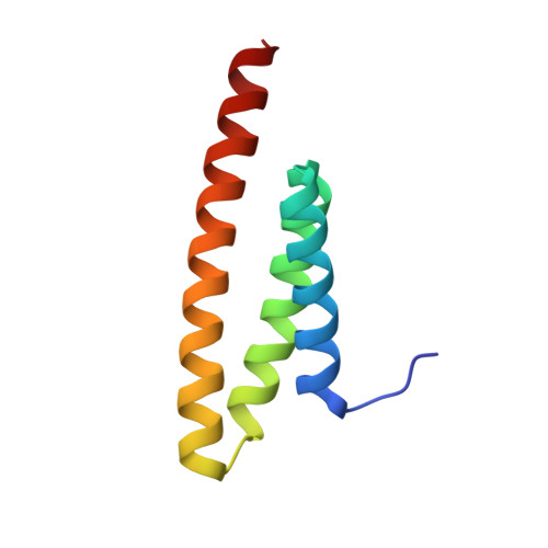
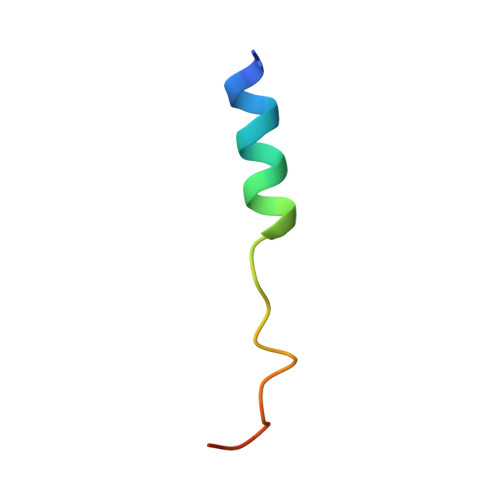
    The endosomal sorting complex required for transport (ESCRT) facilitates roles in membrane remodeling, such as multivesicular body biogenesis, enveloped virus budding and cell division. In yeast, Vps4 plays a crucial role in intraluminal vesicle formation by disassembling ESCRT proteins. Vps4 is recruited by ESCRT-III proteins to the endosomal membrane through the interaction between the microtubule interacting and trafficking (MIT) domain of Vps4 and the C-terminal MIT-interacting motif (MIM) of ESCRT-III proteins. Here, we have determined the crystal structure of Vps4-MIT in a complex with Vps20, a member of ESCRT-III, and revealed that Vps20 adopts a unique MIM2 conformation. Based on structural comparisons with other known MIM2s, we have refined the consensus sequence of MIM2. We have shown that another ESCRT-III protein, Ist1, binds to Vps4-MIT via its C-terminal MIM1 with higher affinity than Vps2, but lacks MIM2 by surface plasmon resonance. Surprisingly, the Ist1 MIM1 competed with the MIM2 of Vfa1, a regulator of Vps4, for binding to Vps4-MIT, even though these MIMs bind in non-overlapping sites on the MIT. These findings provide insight into the allosteric recognition of MIMs of ESCRT-III by Vps4 and also the regulation of ESCRT machinery at the last step of membrane remodeling.


  • Organizational Affiliation

    Faculty of Pharmaceutical Sciences, University of Toyama, 2630 Sugitani, Toyama 930-0194, Japan.


Macromolecules
Find similar proteins by:  (by identity cutoff)  |  3D Structure
Entity ID: 1
MoleculeChains Sequence LengthOrganismDetailsImage
VACUOLAR PROTEIN SORTING-ASSOCIATED PROTEIN 4
A, B
83Saccharomyces cerevisiaeMutation(s): 0 
UniProt
Find proteins for P52917 (Saccharomyces cerevisiae (strain ATCC 204508 / S288c))
Explore P52917 
Go to UniProtKB:  P52917
Entity Groups  
Sequence Clusters30% Identity50% Identity70% Identity90% Identity95% Identity100% Identity
UniProt GroupP52917
Sequence Annotations
Expand
  • Reference Sequence
Find similar proteins by:  (by identity cutoff)  |  3D Structure
Entity ID: 2
MoleculeChains Sequence LengthOrganismDetailsImage
VACUOLAR PROTEIN SORTING-ASSOCIATED PROTEIN 20
C, D
27Saccharomyces cerevisiaeMutation(s): 0 
UniProt
Find proteins for Q04272 (Saccharomyces cerevisiae (strain ATCC 204508 / S288c))
Explore Q04272 
Go to UniProtKB:  Q04272
Entity Groups  
Sequence Clusters30% Identity50% Identity70% Identity90% Identity95% Identity100% Identity
UniProt GroupQ04272
Sequence Annotations
Expand
  • Reference Sequence
Experimental Data & Validation

Experimental Data

  • Method: X-RAY DIFFRACTION
  • Resolution: 1.97 Å
  • R-Value Free: 0.233 
  • R-Value Work: 0.183 
  • R-Value Observed: 0.186 
  • Space Group: P 21 21 21
Unit Cell:
Length ( Å )Angle ( ˚ )
a = 48.14α = 90
b = 53.89β = 90
c = 70.17γ = 90
Software Package:
Software NamePurpose
PHENIXrefinement
MOSFLMdata reduction
SCALAdata scaling
PHASERphasing

Structure Validation

View Full Validation Report


Entry History 

Deposition Data

Revision History  (Full details and data files)

  • Version 1.0: 2016-04-27
    Type: Initial release
  • Version 1.1: 2016-06-08
    Changes: Database references
  • Version 1.2: 2024-01-10
    Changes: Data collection, Database references, Other, Refinement description