5FUL

Crystal Structure of Mus musculus Protein Arginine Methyltransferase 2 with SAH


Experimental Data Snapshot

  • Method: X-RAY DIFFRACTION
  • Resolution: 1.89 Å
  • R-Value Free: 0.192 
  • R-Value Work: 0.166 
  • R-Value Observed: 0.167 

wwPDB Validation   3D Report Full Report


Ligand Structure Quality Assessment 


This is version 1.4 of the entry. See complete history


Literature

Structural studies of protein arginine methyltransferase 2 reveal its interactions with potential substrates and inhibitors.

Cura, V.Marechal, N.Troffer-Charlier, N.Strub, J.M.van Haren, M.J.Martin, N.I.Cianferani, S.Bonnefond, L.Cavarelli, J.

(2017) FEBS J 284: 77-96

  • DOI: https://doi.org/10.1111/febs.13953
  • Primary Citation of Related Structures:  
    5FUB, 5FUL, 5FWA, 5FWD, 5G02, 5JMQ, 5K8V

  • PubMed Abstract: 

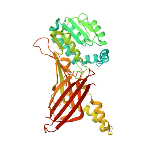
    PRMT2 is the less-characterized member of the protein arginine methyltransferase family in terms of structure, activity, and cellular functions. PRMT2 is a modular protein containing a catalytic Ado-Met-binding domain and unique Src homology 3 domain that binds proteins with proline-rich motifs. PRMT2 is involved in a variety of cellular processes and has diverse roles in transcriptional regulation through different mechanisms depending on its binding partners. PRMT2 has been demonstrated to have weak methyltransferase activity on a histone H4 substrate, but its optimal substrates have not yet been identified. To obtain insights into the function and activity of PRMT2, we solve several crystal structures of PRMT2 from two homologs (zebrafish and mouse) in complex with either the methylation product S-adenosyl-L-homocysteine or other compounds including the first synthetic PRMT2 inhibitor (Cp1) studied so far. We reveal that the N-terminal-containing SH3 module is disordered in the full-length crystal structures, and highlights idiosyncratic features of the PRMT2 active site. We identify a new nonhistone protein substrate belonging to the serine-/arginine-rich protein family which interacts with PRMT2 and we characterize six methylation sites by mass spectrometry. To better understand structural basis for Cp1 binding, we also solve the structure of the complex PRMT4:Cp1. We compare the inhibitor-protein interactions occurring in the PRMT2 and PRMT4 complex crystal structures and show that this compound inhibits efficiently PRMT2. These results are a first step toward a better understanding of PRMT2 substrate recognition and may accelerate the development of structure-based drug design of PRMT2 inhibitors.


  • Organizational Affiliation

    Department of Integrated Structural Biology, Institut de Génétique et de Biologie Moléculaire et Cellulaire (IGBMC), CNRS UMR7104, INSERM U596, Université de Strasbourg, Illkirch, France.


Macromolecules
Find similar proteins by:  (by identity cutoff)  |  3D Structure
Entity ID: 1
MoleculeChains Sequence LengthOrganismDetailsImage
PROTEIN ARGININE N-METHYLTRANSFERASE 2446Mus musculusMutation(s): 1 
EC: 2.1.1.125
UniProt
Find proteins for Q3UKX1 (Mus musculus)
Explore Q3UKX1 
Go to UniProtKB:  Q3UKX1
Entity Groups  
Sequence Clusters30% Identity50% Identity70% Identity90% Identity95% Identity100% Identity
UniProt GroupQ3UKX1
Sequence Annotations
Expand
  • Reference Sequence
Small Molecules
Ligands 5 Unique
IDChains Name / Formula / InChI Key2D Diagram3D Interactions
SAH
Query on SAH

Download Ideal Coordinates CCD File 
B [auth A]S-ADENOSYL-L-HOMOCYSTEINE
C14 H20 N6 O5 S
ZJUKTBDSGOFHSH-WFMPWKQPSA-N
PG4
Query on PG4

Download Ideal Coordinates CCD File 
E [auth A]TETRAETHYLENE GLYCOL
C8 H18 O5
UWHCKJMYHZGTIT-UHFFFAOYSA-N
EDO
Query on EDO

Download Ideal Coordinates CCD File 
G [auth A],
H [auth A]
1,2-ETHANEDIOL
C2 H6 O2
LYCAIKOWRPUZTN-UHFFFAOYSA-N
CA
Query on CA

Download Ideal Coordinates CCD File 
C [auth A],
D [auth A]
CALCIUM ION
Ca
BHPQYMZQTOCNFJ-UHFFFAOYSA-N
CL
Query on CL

Download Ideal Coordinates CCD File 
F [auth A]CHLORIDE ION
Cl
VEXZGXHMUGYJMC-UHFFFAOYSA-M
Binding Affinity Annotations 
IDSourceBinding Affinity
SAH Binding MOAD:  5FUL IC50: 1.83e+4 (nM) from 1 assay(s)
Experimental Data & Validation

Experimental Data

  • Method: X-RAY DIFFRACTION
  • Resolution: 1.89 Å
  • R-Value Free: 0.192 
  • R-Value Work: 0.166 
  • R-Value Observed: 0.167 
  • Space Group: C 2 2 21
Unit Cell:
Length ( Å )Angle ( ˚ )
a = 65.507α = 90
b = 115.414β = 90
c = 132.963γ = 90
Software Package:
Software NamePurpose
PHENIXrefinement
XDSdata reduction
XDSdata scaling
PHASERphasing

Structure Validation

View Full Validation Report


Ligand Structure Quality Assessment 


Entry History 

Deposition Data

Revision History  (Full details and data files)

  • Version 1.0: 2016-11-09
    Type: Initial release
  • Version 1.1: 2016-12-07
    Changes: Database references
  • Version 1.2: 2017-03-01
    Changes: Database references
  • Version 1.3: 2019-04-24
    Changes: Data collection, Source and taxonomy
  • Version 1.4: 2024-01-10
    Changes: Data collection, Database references, Derived calculations, Other, Refinement description