5FT1

Crystal structure of gp37(Dip) from bacteriophage phiKZ bound to RNase E of Pseudomonas aeruginosa


Experimental Data Snapshot

  • Method: X-RAY DIFFRACTION
  • Resolution: 2.75 Å
  • R-Value Free: 0.293 
  • R-Value Work: 0.263 
  • R-Value Observed: 0.266 

wwPDB Validation   3D Report Full Report


This is version 1.2 of the entry. See complete history


Literature

Structural elucidation of a novel mechanism for the bacteriophage-based inhibition of the RNA degradosome.

Van den Bossche, A.Hardwick, S.W.Ceyssens, P.J.Hendrix, H.Voet, M.Dendooven, T.Bandyra, K.J.De Maeyer, M.Aertsen, A.Noben, J.P.Luisi, B.F.Lavigne, R.

(2016) Elife 5

  • DOI: https://doi.org/10.7554/eLife.16413
  • Primary Citation of Related Structures:  
    5FT0, 5FT1

  • PubMed Abstract: 

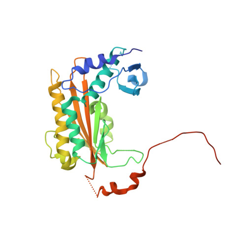
    In all domains of life, the catalysed degradation of RNA facilitates rapid adaptation to changing environmental conditions, while destruction of foreign RNA is an important mechanism to prevent host infection. We have identified a virus-encoded protein termed gp37/Dip, which directly binds and inhibits the RNA degradation machinery of its bacterial host. Encoded by giant phage фKZ, this protein associates with two RNA binding sites of the RNase E component of the Pseudomonas aeruginosa RNA degradosome, occluding them from substrates and resulting in effective inhibition of RNA degradation and processing. The 2.2 Å crystal structure reveals that this novel homo-dimeric protein has no identifiable structural homologues. Our biochemical data indicate that acidic patches on the convex outer surface bind RNase E. Through the activity of Dip, фKZ has evolved a unique mechanism to down regulate a key metabolic process of its host to allow accumulation of viral RNA in infected cells.


  • Organizational Affiliation

    Laboratory of Gene Technology, KU Leuven, Leuven, Belgium.


Macromolecules
Find similar proteins by:  (by identity cutoff)  |  3D Structure
Entity ID: 1
MoleculeChains Sequence LengthOrganismDetailsImage
GP37
A, B, E, G, I
A, B, E, G, I, K
281Phikzvirus phiKZMutation(s): 0 
UniProt
Find proteins for Q8SDC5 (Pseudomonas phage phiKZ)
Explore Q8SDC5 
Go to UniProtKB:  Q8SDC5
Entity Groups  
Sequence Clusters30% Identity50% Identity70% Identity90% Identity95% Identity100% Identity
UniProt GroupQ8SDC5
Sequence Annotations
Expand
  • Reference Sequence

Find similar proteins by:  Sequence   |   3D Structure  

Entity ID: 2
MoleculeChains Sequence LengthOrganismDetailsImage
RIBONUCLEASE E
C, D, F, H, J
C, D, F, H, J, L
20Pseudomonas aeruginosaMutation(s): 0 
EC: 3.1.26.12
UniProt
Find proteins for Q9HZM8 (Pseudomonas aeruginosa (strain ATCC 15692 / DSM 22644 / CIP 104116 / JCM 14847 / LMG 12228 / 1C / PRS 101 / PAO1))
Explore Q9HZM8 
Go to UniProtKB:  Q9HZM8
Entity Groups  
Sequence Clusters30% Identity50% Identity70% Identity90% Identity95% Identity100% Identity
UniProt GroupQ9HZM8
Sequence Annotations
Expand
  • Reference Sequence
Experimental Data & Validation

Experimental Data

  • Method: X-RAY DIFFRACTION
  • Resolution: 2.75 Å
  • R-Value Free: 0.293 
  • R-Value Work: 0.263 
  • R-Value Observed: 0.266 
  • Space Group: P 1
Unit Cell:
Length ( Å )Angle ( ˚ )
a = 84.61α = 107.86
b = 84.65β = 107.88
c = 84.6γ = 107.9
Software Package:
Software NamePurpose
PHENIXrefinement
MOSFLMdata reduction
Aimlessdata scaling
PHASERphasing

Structure Validation

View Full Validation Report


Entry History 

Revision History  (Full details and data files)

  • Version 1.0: 2016-08-03
    Type: Initial release
  • Version 1.1: 2017-03-22
    Changes: Database references
  • Version 1.2: 2024-01-10
    Changes: Data collection, Database references, Other, Refinement description