5FQD

Structural basis of Lenalidomide induced CK1a degradation by the crl4crbn ubiquitin ligase


Experimental Data Snapshot

  • Method: X-RAY DIFFRACTION
  • Resolution: 2.45 Å
  • R-Value Free: 0.210 
  • R-Value Work: 0.181 
  • R-Value Observed: 0.182 

wwPDB Validation   3D Report Full Report


Ligand Structure Quality Assessment 


This is version 1.4 of the entry. See complete history


Literature

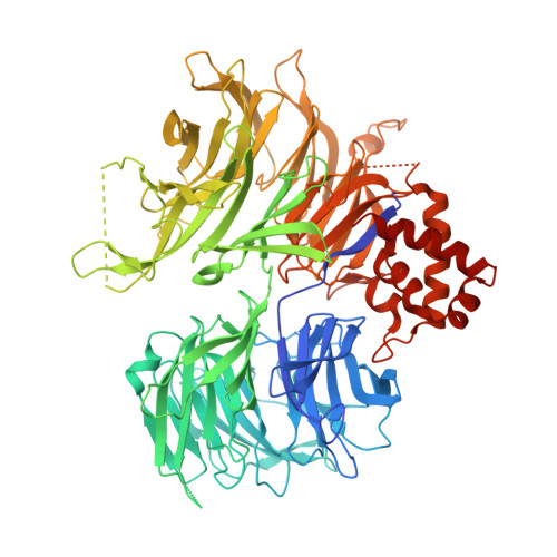
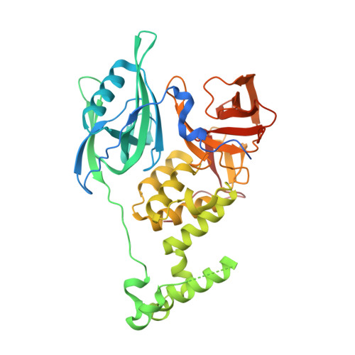
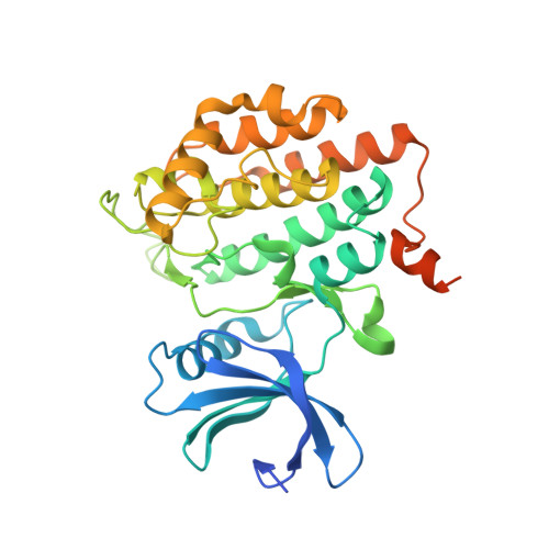
Structural Basis of Lenalidomide-Induced Ck1Alpha Degradation by the Crl4(Crbn) Ubiquitin Ligase.

Petzold, G.Fischer, E.S.Thoma, N.H.

(2016) Nature 532: 127

  • DOI: https://doi.org/10.1038/nature16979
  • Primary Citation of Related Structures:  
    5FQD

  • PubMed Abstract: 

    Thalidomide and its derivatives, lenalidomide and pomalidomide, are immune modulatory drugs (IMiDs) used in the treatment of haematologic malignancies. IMiDs bind CRBN, the substrate receptor of the CUL4-RBX1-DDB1-CRBN (also known as CRL4(CRBN)) E3 ubiquitin ligase, and inhibit ubiquitination of endogenous CRL4(CRBN) substrates. Unexpectedly, IMiDs also repurpose the ligase to target new proteins for degradation. Lenalidomide induces degradation of the lymphoid transcription factors Ikaros and Aiolos (also known as IKZF1 and IKZF3), and casein kinase 1α (CK1α), which contributes to its clinical efficacy in the treatment of multiple myeloma and 5q-deletion associated myelodysplastic syndrome (del(5q) MDS), respectively. How lenalidomide alters the specificity of the ligase to degrade these proteins remains elusive. Here we present the 2.45 Å crystal structure of DDB1-CRBN bound to lenalidomide and CK1α. CRBN and lenalidomide jointly provide the binding interface for a CK1α β-hairpin-loop located in the kinase N-lobe. We show that CK1α binding to CRL4(CRBN) is strictly dependent on the presence of an IMiD. Binding of IKZF1 to CRBN similarly requires the compound and both, IKZF1 and CK1α, use a related binding mode. Our study provides a mechanistic explanation for the selective efficacy of lenalidomide in del(5q) MDS therapy. We anticipate that high-affinity protein-protein interactions induced by small molecules will provide opportunities for drug development, particularly for targeted protein degradation.


  • Organizational Affiliation

    Friedrich Miescher Institute for Biomedical Research, Maulbeerstrasse 66, CH-4058 Basel, Switzerland.


Macromolecules
Find similar proteins by:  (by identity cutoff)  |  3D Structure
Entity ID: 1
MoleculeChains Sequence LengthOrganismDetailsImage
DNA DAMAGE-BINDING PROTEIN 1
A, D
856Homo sapiensMutation(s): 0 
EC: 6.3.2.19
UniProt & NIH Common Fund Data Resources
Find proteins for Q16531 (Homo sapiens)
Explore Q16531 
Go to UniProtKB:  Q16531
PHAROS:  Q16531
GTEx:  ENSG00000167986 
Entity Groups  
Sequence Clusters30% Identity50% Identity70% Identity90% Identity95% Identity100% Identity
UniProt GroupQ16531
Sequence Annotations
Expand
  • Reference Sequence
Find similar proteins by:  (by identity cutoff)  |  3D Structure
Entity ID: 2
MoleculeChains Sequence LengthOrganismDetailsImage
PROTEIN CEREBLON
B, E
426Homo sapiensMutation(s): 0 
UniProt & NIH Common Fund Data Resources
Find proteins for Q96SW2 (Homo sapiens)
Explore Q96SW2 
Go to UniProtKB:  Q96SW2
PHAROS:  Q96SW2
GTEx:  ENSG00000113851 
Entity Groups  
Sequence Clusters30% Identity50% Identity70% Identity90% Identity95% Identity100% Identity
UniProt GroupQ96SW2
Sequence Annotations
Expand
  • Reference Sequence
Find similar proteins by:  (by identity cutoff)  |  3D Structure
Entity ID: 3
MoleculeChains Sequence LengthOrganismDetailsImage
CASEIN KINASE I ISOFORM ALPHA
C, F
341Homo sapiensMutation(s): 0 
UniProt & NIH Common Fund Data Resources
Find proteins for P48729 (Homo sapiens)
Explore P48729 
Go to UniProtKB:  P48729
PHAROS:  P48729
GTEx:  ENSG00000113712 
Entity Groups  
Sequence Clusters30% Identity50% Identity70% Identity90% Identity95% Identity100% Identity
UniProt GroupP48729
Sequence Annotations
Expand
  • Reference Sequence
Small Molecules
Binding Affinity Annotations 
IDSourceBinding Affinity
LVY BindingDB:  5FQD Ki: min: 1490, max: 1.63e+4 (nM) from 2 assay(s)
IC50: min: 1500, max: 3.78e+4 (nM) from 4 assay(s)
Binding MOAD:  5FQD Kd: 242 (nM) from 1 assay(s)
Experimental Data & Validation

Experimental Data

  • Method: X-RAY DIFFRACTION
  • Resolution: 2.45 Å
  • R-Value Free: 0.210 
  • R-Value Work: 0.181 
  • R-Value Observed: 0.182 
  • Space Group: P 1
Unit Cell:
Length ( Å )Angle ( ˚ )
a = 87.97α = 106.02
b = 109.93β = 93.19
c = 112.4γ = 101.63
Software Package:
Software NamePurpose
BUSTERrefinement
XDSdata reduction
XSCALEdata scaling
PHASERphasing

Structure Validation

View Full Validation Report


Ligand Structure Quality Assessment 


Entry History 

Deposition Data

Revision History  (Full details and data files)

  • Version 1.0: 2016-02-24
    Type: Initial release
  • Version 1.1: 2016-03-09
    Changes: Database references
  • Version 1.2: 2016-04-13
    Changes: Database references
  • Version 1.3: 2019-04-03
    Changes: Data collection, Other, Source and taxonomy
  • Version 1.4: 2024-01-10
    Changes: Data collection, Database references, Derived calculations, Other, Refinement description