Structural basis for nutrient acquisition by dominant members of the human gut microbiota.
Glenwright, A.J., Pothula, K.R., Bhamidimarri, S.P., Chorev, D.S., Basle, A., Firbank, S.J., Zheng, H., Robinson, C.V., Winterhalter, M., Kleinekathofer, U., Bolam, D.N., van den Berg, B.(2017) Nature 541: 407-411
- PubMed: 28077872
- DOI: https://doi.org/10.1038/nature20828
- Primary Citation of Related Structures:
5FQ3, 5FQ4, 5FQ6, 5FQ7, 5FQ8, 5LX8, 5T3R, 5T4Y - PubMed Abstract:
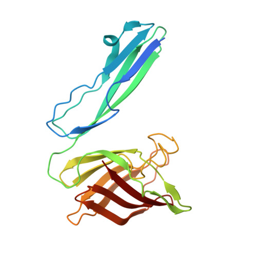
The human large intestine is populated by a high density of microorganisms, collectively termed the colonic microbiota, which has an important role in human health and nutrition. The survival of microbiota members from the dominant Gram-negative phylum Bacteroidetes depends on their ability to degrade dietary glycans that cannot be metabolized by the host. The genes encoding proteins involved in the degradation of specific glycans are organized into co-regulated polysaccharide utilization loci, with the archetypal locus sus (for starch utilisation system) encoding seven proteins, SusA-SusG. Glycan degradation mainly occurs intracellularly and depends on the import of oligosaccharides by an outer membrane protein complex composed of an extracellular SusD-like lipoprotein and an integral membrane SusC-like TonB-dependent transporter. The presence of the partner SusD-like lipoprotein is the major feature that distinguishes SusC-like proteins from previously characterized TonB-dependent transporters. Many sequenced gut Bacteroides spp. encode over 100 SusCD pairs, of which the majority have unknown functions and substrate specificities. The mechanism by which extracellular substrate binding by SusD proteins is coupled to outer membrane passage through their cognate SusC transporter is unknown. Here we present X-ray crystal structures of two functionally distinct SusCD complexes purified from Bacteroides thetaiotaomicron and derive a general model for substrate translocation. The SusC transporters form homodimers, with each β-barrel protomer tightly capped by SusD. Ligands are bound at the SusC-SusD interface in a large solvent-excluded cavity. Molecular dynamics simulations and single-channel electrophysiology reveal a 'pedal bin' mechanism, in which SusD moves away from SusC in a hinge-like fashion in the absence of ligand to expose the substrate-binding site to the extracellular milieu. These data provide mechanistic insights into outer membrane nutrient import by members of the microbiota, an area of major importance for understanding human-microbiota symbiosis.
Organizational Affiliation:
Institute for Cell and Molecular Biosciences, The Medical School, Newcastle University, Newcastle upon Tyne NE2 4HH, UK.