


2.1 Angstrom Crystal Structure of Pro Cathepsin B S9 from Trypanosoma Congolense
Sevajol, M., Biteau, N., Baltz, T., Franzetti, B., Vellieux, F.M.D.(null) Ph D Thesis
Experimental Data Snapshot
wwPDB Validation 3D Report Full Report

(null) Ph D Thesis
Entity ID: 1 | |||||
|---|---|---|---|---|---|
| Molecule | Chains | Sequence Length | Organism | Details | Image |
| PRO CATHEPSIN B S9 | 321 | Trypanosoma congolense | Mutation(s): 2 EC: 3.4.22.1 | 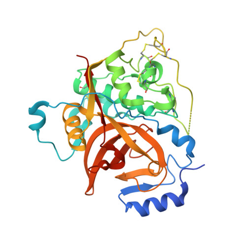 | |
UniProt | |||||
Find proteins for B2C331 (Trypanosoma congolense) Explore B2C331 Go to UniProtKB: B2C331 | |||||
Entity Groups | |||||
| Sequence Clusters | 30% Identity50% Identity70% Identity90% Identity95% Identity100% Identity | ||||
| UniProt Group | B2C331 | ||||
Sequence AnnotationsExpand | |||||
| |||||
| Length ( Å ) | Angle ( ˚ ) |
|---|---|
| a = 128.79 | α = 90 |
| b = 223.22 | β = 90 |
| c = 102.89 | γ = 90 |
| Software Name | Purpose |
|---|---|
| PHENIX | refinement |
| XDS | data reduction |
| XSCALE | data scaling |
RCSB PDB (citation) is hosted by
RCSB PDB is a member of the

