5FDK

Crystal structure of RecU(D88N) in complex with palindromic DNA duplex


Experimental Data Snapshot

  • Method: X-RAY DIFFRACTION
  • Resolution: 3.21 Å
  • R-Value Free: 0.329 
  • R-Value Work: 0.256 
  • R-Value Observed: 0.260 

wwPDB Validation   3D Report Full Report


This is version 1.2 of the entry. See complete history


Literature

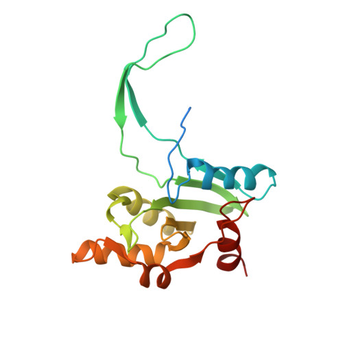
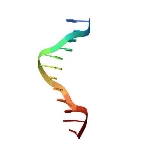
Structural insights into dynamics of RecU-HJ complex formation elucidates key role of NTR and stalk region toward formation of reactive state.

Khavnekar, S.Dantu, S.C.Sedelnikova, S.Ayora, S.Rafferty, J.Kale, A.

(2017) Nucleic Acids Res 45: 975-986

  • DOI: https://doi.org/10.1093/nar/gkw1165
  • Primary Citation of Related Structures:  
    5FDK

  • PubMed Abstract: 

    Holliday junction (HJ) resolving enzyme RecU is involved in DNA repair and recombination. We have determined the crystal structure of inactive mutant (D88N) of RecU from Bacillus subtilis in complex with a 12 base palindromic DNA fragment at a resolution of 3.2 Å. This structure shows the stalk region and the essential N-terminal region (NTR) previously unseen in our DNA unbound structure. The flexible nature of the NTR in solution was confirmed using SAXS. Thermofluor studies performed to assess the stability of RecU in complex with the arms of an HJ indicate that it confers stability. Further, we performed molecular dynamics (MD) simulations of wild type and an NTR deletion variant of RecU, with and without HJ. The NTR is observed to be highly flexible in simulations of the unbound RecU, in agreement with SAXS observations. These simulations revealed domain dynamics of RecU and their role in the formation of complex with HJ. The MD simulations also elucidate key roles of the NTR, stalk region, and breathing motion of RecU in the formation of the reactive state.


  • Organizational Affiliation

    UM-DAE Centre for Excellence in Basic Science, University of Mumbai, Vidhyanagari Campus, Mumbai 400098, India.


Macromolecules

Find similar proteins by:  (by identity cutoff)  |  3D Structure
Entity ID: 1
MoleculeChains Sequence LengthOrganismDetailsImage
Holliday junction resolvase RecU
A, B, C, D
199Bacillus subtilisMutation(s): 1 
Gene Names: recUprfAyppBBSU22310
EC: 3.1.22
UniProt
Find proteins for P39792 (Bacillus subtilis (strain 168))
Explore P39792 
Go to UniProtKB:  P39792
Entity Groups  
Sequence Clusters30% Identity50% Identity70% Identity90% Identity95% Identity100% Identity
UniProt GroupP39792
Sequence Annotations
Expand
  • Reference Sequence

Find similar nucleic acids by:  Sequence   |   3D Structure  

Entity ID: 2
MoleculeChains LengthOrganismImage
palindromic DNA
E, F, G, H
12synthetic construct
Sequence Annotations
Expand
  • Reference Sequence
Experimental Data & Validation

Experimental Data

  • Method: X-RAY DIFFRACTION
  • Resolution: 3.21 Å
  • R-Value Free: 0.329 
  • R-Value Work: 0.256 
  • R-Value Observed: 0.260 
  • Space Group: H 3 2
Unit Cell:
Length ( Å )Angle ( ˚ )
a = 144.447α = 90
b = 144.447β = 90
c = 310.343γ = 120
Software Package:
Software NamePurpose
SCALAdata scaling
PHASERphasing
PHENIXrefinement
PDB_EXTRACTdata extraction
XDSdata reduction
Cootmodel building

Structure Validation

View Full Validation Report


Entry History 

Deposition Data

Revision History  (Full details and data files)

  • Version 1.0: 2016-12-07
    Type: Initial release
  • Version 1.1: 2016-12-14
    Changes: Database references
  • Version 1.2: 2017-02-08
    Changes: Database references