5FB6

Room-temperature macromolecular crystallography using a micro-patterned silicon chip with minimal background scattering


Experimental Data Snapshot

  • Method: X-RAY DIFFRACTION
  • Resolution: 1.90 Å
  • R-Value Free: 0.173 
  • R-Value Work: 0.157 
  • R-Value Observed: 0.159 

wwPDB Validation   3D Report Full Report


This is version 1.2 of the entry. See complete history


Literature

Room-temperature macromolecular crystallography using a micro-patterned silicon chip with minimal background scattering.

Roedig, P.Duman, R.Sanchez-Weatherby, J.Vartiainen, I.Burkhardt, A.Warmer, M.David, C.Wagner, A.Meents, A.

(2016) J Appl Crystallogr 49: 968-975

  • DOI: https://doi.org/10.1107/S1600576716006348
  • Primary Citation of Related Structures:  
    5FB6

  • PubMed Abstract: 

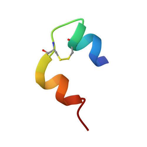
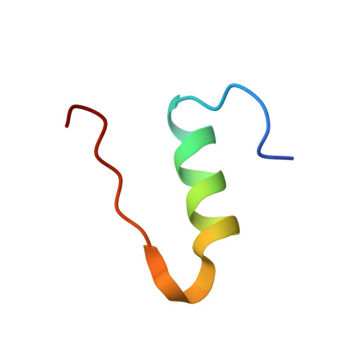
    Recent success at X-ray free-electron lasers has led to serial crystallography experiments staging a comeback at synchrotron sources as well. With crystal lifetimes typically in the millisecond range and the latest-generation detector technologies with high framing rates up to 1 kHz, fast sample exchange has become the bottleneck for such experiments. A micro-patterned chip has been developed from single-crystalline silicon, which acts as a sample holder for up to several thousand microcrystals at a very low background level. The crystals can be easily loaded onto the chip and excess mother liquor can be efficiently removed. Dehydration of the crystals is prevented by keeping them in a stream of humidified air during data collection. Further sealing of the sample holder, for example with Kapton, is not required. Room-temperature data collection from insulin crystals loaded onto the chip proves the applicability of the chip for macromolecular crystallography. Subsequent structure refinements reveal no radiation-damage-induced structural changes for insulin crystals up to a dose of 565.6 kGy, even though the total diffraction power of the crystals has on average decreased to 19.1% of its initial value for the same dose. A decay of the diffracting power by half is observed for a dose of D 1/2 = 147.5 ± 19.1 kGy, which is about 1/300 of the dose before crystals show a similar decay at cryogenic temperatures.


  • Organizational Affiliation

    Deutsches Elektronen-Synchrotron DESY, Photon Science, Notkestrasse 85, Hamburg 22607, Germany.


Macromolecules

Find similar proteins by:  Sequence   |   3D Structure  

Entity ID: 1
MoleculeChains Sequence LengthOrganismDetailsImage
Insulin Chain A21Sus scrofaMutation(s): 0 
UniProt
Find proteins for P01315 (Sus scrofa)
Explore P01315 
Go to UniProtKB:  P01315
Entity Groups  
Sequence Clusters30% Identity50% Identity70% Identity90% Identity95% Identity100% Identity
UniProt GroupP01315
Sequence Annotations
Expand
  • Reference Sequence
Find similar proteins by:  (by identity cutoff)  |  3D Structure
Entity ID: 2
MoleculeChains Sequence LengthOrganismDetailsImage
Insulin Chain B30Sus scrofaMutation(s): 0 
UniProt
Find proteins for P01315 (Sus scrofa)
Explore P01315 
Go to UniProtKB:  P01315
Entity Groups  
Sequence Clusters30% Identity50% Identity70% Identity90% Identity95% Identity100% Identity
UniProt GroupP01315
Sequence Annotations
Expand
  • Reference Sequence
Experimental Data & Validation

Experimental Data

  • Method: X-RAY DIFFRACTION
  • Resolution: 1.90 Å
  • R-Value Free: 0.173 
  • R-Value Work: 0.157 
  • R-Value Observed: 0.159 
  • Space Group: I 21 3
Unit Cell:
Length ( Å )Angle ( ˚ )
a = 78.82α = 90
b = 78.82β = 90
c = 78.82γ = 90
Software Package:
Software NamePurpose
PHENIXrefinement
XSCALEdata scaling
XDSdata reduction
PHASERphasing

Structure Validation

View Full Validation Report


Entry History & Funding Information

Deposition Data


Funding OrganizationLocationGrant Number
European CommunitySwitzerland290605
Helmholtz AssociationGermanyVH-VI-403

Revision History  (Full details and data files)

  • Version 1.0: 2016-06-15
    Type: Initial release
  • Version 1.1: 2016-06-22
    Changes: Database references
  • Version 1.2: 2024-01-10
    Changes: Data collection, Database references, Refinement description