5FB3

Structure of glycerophosphate dehydrogenase in complex with NADPH


Experimental Data Snapshot

  • Method: X-RAY DIFFRACTION
  • Resolution: 2.45 Å
  • R-Value Free: 0.260 
  • R-Value Work: 0.207 
  • R-Value Observed: 0.210 

wwPDB Validation   3D Report Full Report


Ligand Structure Quality Assessment 


This is version 1.3 of the entry. See complete history


Literature

Unique coenzyme binding mode of hyperthermophilic archaeal sn-glycerol-1-phosphate dehydrogenase from Pyrobaculum calidifontis

Hayashi, J.Yamamoto, K.Yoneda, K.Ohshima, T.Sakuraba, H.

(2016) Proteins 84: 1786-1796

  • DOI: https://doi.org/10.1002/prot.25161
  • Primary Citation of Related Structures:  
    5FB3

  • PubMed Abstract: 

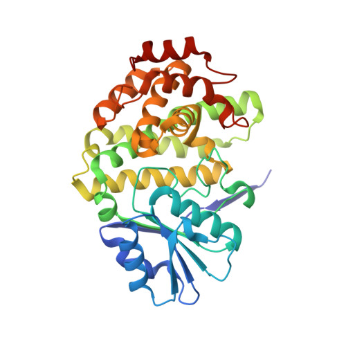
    A gene encoding an sn-glycerol-1-phosphate dehydrogenase (G1PDH) was identified in the hyperthermophilic archaeon Pyrobaculum calidifontis. The gene was overexpressed in Escherichia coli, and its product was purified and characterized. In contrast to conventional G1PDHs, the expressed enzyme showed strong preference for NADH: the reaction rate (V max ) with NADPH was only 2.4% of that with NADH. The crystal structure of the enzyme was determined at a resolution of 2.45 Å. The asymmetric unit consisted of one homohexamer. Refinement of the structure and HPLC analysis showed the presence of the bound cofactor NADPH in subunits D, E, and F, even though it was not added in the crystallization procedure. The phosphate group at C2' of the adenine ribose of NADPH is tightly held through the five biased hydrogen bonds with Ser40 and Thr42. In comparison with the known G1PDH structure, the NADPH molecule was observed to be pushed away from the normal coenzyme binding site. Interestingly, the S40A/T42A double mutant enzyme acquired much higher reactivity than the wild-type enzyme with NADPH, which suggests that the biased interactions around the C2'-phosphate group make NADPH binding insufficient for catalysis. Our results provide a unique structural basis for coenzyme preference in NAD(P)-dependent dehydrogenases. Proteins 2016; 84:1786-1796. © 2016 Wiley Periodicals, Inc.


  • Organizational Affiliation

    Department of Applied Biological Science, Faculty of Agriculture, Kagawa University, 2393 Ikenobe, Miki-cho, Kita-gun, Kagawa, 761-0795, Japan.


Macromolecules
Find similar proteins by:  (by identity cutoff)  |  3D Structure
Entity ID: 1
MoleculeChains Sequence LengthOrganismDetailsImage
Glycerol-1-phosphate dehydrogenase [NAD(P)+]
A, B, C, D, E
A, B, C, D, E, F
338Pyrobaculum calidifontis JCM 11548Mutation(s): 0 
Gene Names: egsAPcal_0566
EC: 1.1.1.261
UniProt
Find proteins for A3MTM6 (Pyrobaculum calidifontis (strain DSM 21063 / JCM 11548 / VA1))
Explore A3MTM6 
Go to UniProtKB:  A3MTM6
Entity Groups  
Sequence Clusters30% Identity50% Identity70% Identity90% Identity95% Identity100% Identity
UniProt GroupA3MTM6
Sequence Annotations
Expand
  • Reference Sequence
Small Molecules
Ligands 4 Unique
IDChains Name / Formula / InChI Key2D Diagram3D Interactions
NDP
Query on NDP

Download Ideal Coordinates CCD File 
R [auth D],
U [auth E],
Y [auth F]
NADPH DIHYDRO-NICOTINAMIDE-ADENINE-DINUCLEOTIDE PHOSPHATE
C21 H30 N7 O17 P3
ACFIXJIJDZMPPO-NNYOXOHSSA-N
PPV
Query on PPV

Download Ideal Coordinates CCD File 
N [auth B]PYROPHOSPHATE
H4 O7 P2
XPPKVPWEQAFLFU-UHFFFAOYSA-N
SO4
Query on SO4

Download Ideal Coordinates CCD File 
AA [auth F]
BA [auth F]
H [auth A]
I [auth A]
J [auth A]
AA [auth F],
BA [auth F],
H [auth A],
I [auth A],
J [auth A],
L [auth B],
M [auth B],
P [auth C],
Q [auth C],
T [auth D],
W [auth E],
X [auth F]
SULFATE ION
O4 S
QAOWNCQODCNURD-UHFFFAOYSA-L
ZN
Query on ZN

Download Ideal Coordinates CCD File 
G [auth A]
K [auth B]
O [auth C]
S [auth D]
V [auth E]
G [auth A],
K [auth B],
O [auth C],
S [auth D],
V [auth E],
Z [auth F]
ZINC ION
Zn
PTFCDOFLOPIGGS-UHFFFAOYSA-N
Experimental Data & Validation

Experimental Data

  • Method: X-RAY DIFFRACTION
  • Resolution: 2.45 Å
  • R-Value Free: 0.260 
  • R-Value Work: 0.207 
  • R-Value Observed: 0.210 
  • Space Group: P 21 21 21
Unit Cell:
Length ( Å )Angle ( ˚ )
a = 102.182α = 90
b = 123.335β = 90
c = 166.724γ = 90
Software Package:
Software NamePurpose
HKL-2000data collection
HKL-2000data scaling
REFMACrefinement
PDB_EXTRACTdata extraction
BUCCANEERmodel building

Structure Validation

View Full Validation Report


Ligand Structure Quality Assessment 


Entry History 

Deposition Data

Revision History  (Full details and data files)

  • Version 1.0: 2016-10-12
    Type: Initial release
  • Version 1.1: 2016-12-07
    Changes: Database references
  • Version 1.2: 2020-02-19
    Changes: Data collection, Database references
  • Version 1.3: 2024-03-20
    Changes: Data collection, Database references