5FB1

Crystal Structure of a PHD finger bound to histone H3 K9me3 peptide


Experimental Data Snapshot

  • Method: X-RAY DIFFRACTION
  • Resolution: 2.10 Å
  • R-Value Free: 0.220 
  • R-Value Work: 0.167 
  • R-Value Observed: 0.169 

wwPDB Validation   3D Report Full Report


This is version 1.3 of the entry. See complete history


Literature

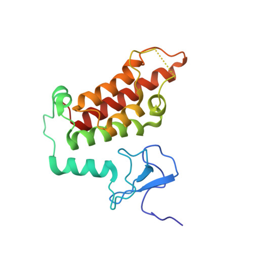
Multifaceted Histone H3 Methylation and Phosphorylation Readout by the Plant Homeodomain Finger of Human Nuclear Antigen Sp100C

Zhang, X.Zhao, D.Xiong, X.He, Z.Li, H.

(2016) J Biol Chem 291: 12786-12798

  • DOI: https://doi.org/10.1074/jbc.M116.721159
  • Primary Citation of Related Structures:  
    5FB0, 5FB1

  • PubMed Abstract: 

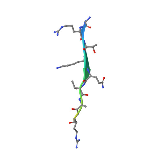
    The decoding of histone post-translational modifications by chromatin-binding modules ("readers") constitutes one major mechanism of epigenetic regulation. Nuclear antigen Sp100 (SPECKLED, 100 kDa), a constitutive component of the promyelocytic leukemia nuclear bodies, plays key roles in intrinsic immunity and transcriptional repression. Sp100C, a splicing isoform specifically up-regulated upon interferon stimulation, harbors a unique tandem plant homeodomain (PHD) finger and bromodomain at its C terminus. Combining structural, quantitative binding, and cellular co-localization studies, we characterized Sp100C PHD finger as an unmethylated histone H3 Lys(4) (H3K4me0) reader that tolerates histone H3 Thr(3) phosphorylation (H3T3ph), histone H3 Lys(9) trimethylation (H3K9me3), and histone H3 Ser(10) phosphorylation (H3S10ph), hallmarks associated with the mitotic chromosome. In contrast, whereas H3K4me0 reader activity is conserved in Sp140, an Sp100C paralog, the multivalent tolerance of H3T3ph, H3K9me3, and H3S10ph was lost for Sp140. The complex structure determined at 2.1 Å revealed a highly coordinated lysine ϵ-amine recognition sphere formed by an extended N-terminal motif for H3K4me0 readout. Interestingly, reader pocket rigidification by disulfide bond formation enhanced H3K4me0 binding by Sp100C. An additional complex structure solved at 2.7 Å revealed that H3T3ph is recognized by the arginine residue, Arg(713), that is unique to the PHD finger of Sp100C. Consistent with a restrictive cellular role of Sp100C, these results establish a direct chromatin targeting function of Sp100C that may regulate transcriptional gene silencing and promyelocytic leukemia nuclear body-mediated intrinsic immunity in response to interferon stimulation.


  • Organizational Affiliation

    From the Ministry of Education Key Laboratory of Protein Sciences, Beijing Advanced Innovation Center for Structural Biology, Department of Basic Medical Sciences, School of Medicine, Tsinghua University, Beijing 100084 and.


Macromolecules
Find similar proteins by:  (by identity cutoff)  |  3D Structure
Entity ID: 1
MoleculeChains Sequence LengthOrganismDetailsImage
Nuclear autoantigen Sp-100181Homo sapiensMutation(s): 1 
Gene Names: SP100
UniProt & NIH Common Fund Data Resources
Find proteins for P23497 (Homo sapiens)
Explore P23497 
Go to UniProtKB:  P23497
PHAROS:  P23497
GTEx:  ENSG00000067066 
Entity Groups  
Sequence Clusters30% Identity50% Identity70% Identity90% Identity95% Identity100% Identity
UniProt GroupP23497
Sequence Annotations
Expand
  • Reference Sequence

Find similar proteins by:  Sequence   |   3D Structure  

Entity ID: 2
MoleculeChains Sequence LengthOrganismDetailsImage
Peptide from Histone H3B [auth C]15Homo sapiensMutation(s): 0 
UniProt & NIH Common Fund Data Resources
Find proteins for P68431 (Homo sapiens)
Explore P68431 
Go to UniProtKB:  P68431
PHAROS:  P68431
Entity Groups  
Sequence Clusters30% Identity50% Identity70% Identity90% Identity95% Identity100% Identity
UniProt GroupP68431
Sequence Annotations
Expand
  • Reference Sequence
Experimental Data & Validation

Experimental Data

  • Method: X-RAY DIFFRACTION
  • Resolution: 2.10 Å
  • R-Value Free: 0.220 
  • R-Value Work: 0.167 
  • R-Value Observed: 0.169 
  • Space Group: P 21 21 2
Unit Cell:
Length ( Å )Angle ( ˚ )
a = 52.84α = 90
b = 102.964β = 90
c = 44.955γ = 90
Software Package:
Software NamePurpose
HKL-2000data scaling
HKL-2000data reduction
AutoSolphasing
PHENIXrefinement
PDB_EXTRACTdata extraction

Structure Validation

View Full Validation Report


Entry History & Funding Information

Deposition Data


Funding OrganizationLocationGrant Number
Major State Basic Research Development ProgramChina2015CB910503

Revision History  (Full details and data files)

  • Version 1.0: 2016-05-04
    Type: Initial release
  • Version 1.1: 2016-06-22
    Changes: Database references
  • Version 1.2: 2017-10-04
    Changes: Data collection, Database references, Derived calculations
  • Version 1.3: 2024-03-20
    Changes: Data collection, Database references