


5F5Q
Crystal structure of Canavalia virosa lectin in complex with alpha-methyl-mannoside
- PDB DOI: https://doi.org/10.2210/pdb5F5Q/pdb
- Classification: SUGAR BINDING PROTEIN
- Organism(s): Canavalia cathartica
- Mutation(s): No
- Deposited: 2015-12-04 Released: 2016-10-26
Experimental Data Snapshot
- Method: X-RAY DIFFRACTION
- Resolution: 2.52 Å
- R-Value Free: 0.251
- R-Value Work: 0.184
- R-Value Observed: 0.188

This is version 1.4 of the entry. See complete history.

Macromolecules
Find similar proteins by:
(by identity cutoff) | 3D Structure
Entity ID: 1 | |||||
|---|---|---|---|---|---|
| Molecule | Chains | Sequence Length | Organism | Details | Image |
| Concanavalin-A | 237 | Canavalia cathartica | Mutation(s): 0 | 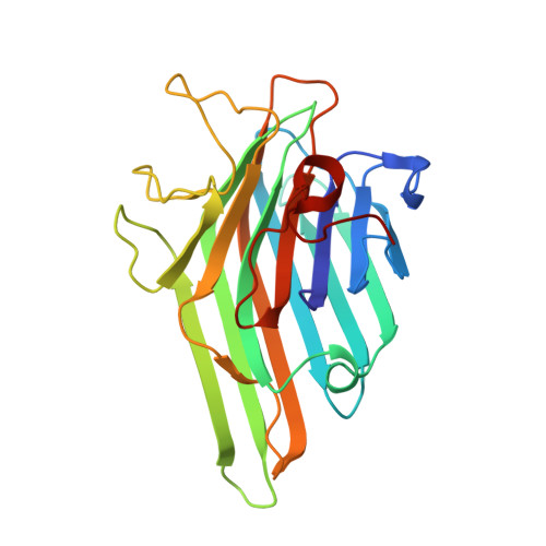 | |
UniProt | |||||
Find proteins for C0HJY1 (Canavalia cathartica) Explore C0HJY1 Go to UniProtKB: C0HJY1 | |||||
Entity Groups | |||||
| Sequence Clusters | 30% Identity50% Identity70% Identity90% Identity95% Identity100% Identity | ||||
| UniProt Group | C0HJY1 | ||||
Sequence AnnotationsExpand | |||||
| |||||
Small Molecules
| Ligands 3 Unique | |||||
|---|---|---|---|---|---|
| ID | Chains | Name / Formula / InChI Key | 2D Diagram | 3D Interactions | |
| MMA Query on MMA | E [auth A], H [auth B] | methyl alpha-D-mannopyranoside C7 H14 O6 HOVAGTYPODGVJG-VEIUFWFVSA-N |  | ||
| MN Query on MN | D [auth A], G [auth B] | MANGANESE (II) ION Mn WAEMQWOKJMHJLA-UHFFFAOYSA-N |  | ||
| CA Query on CA | C [auth A], F [auth B] | CALCIUM ION Ca BHPQYMZQTOCNFJ-UHFFFAOYSA-N |  | ||
Experimental Data & Validation
Experimental Data
- Method: X-RAY DIFFRACTION
- Resolution: 2.52 Å
- R-Value Free: 0.251
- R-Value Work: 0.184
- R-Value Observed: 0.188
- Space Group: P 21 2 21
Unit Cell:
| Length ( Å ) | Angle ( ˚ ) |
|---|---|
| a = 60.25 | α = 90 |
| b = 81.74 | β = 90 |
| c = 86.93 | γ = 90 |
| Software Name | Purpose |
|---|---|
| REFMAC | refinement |
| SCALA | data scaling |
| PDB_EXTRACT | data extraction |
| iMOSFLM | data reduction |
| MOLREP | phasing |
Entry History
Deposition Data
- Released Date: 2016-10-26 Deposition Author(s): Osterne, V.J.S., Silva-Filho, J.C., Pinto-Junior, V.R., Santiago, M.Q., Lossio, C.F., Delatorre, P., Nascimento, K.S., Cavada, B.S.
Revision History (Full details and data files)
- Version 1.0: 2016-10-26
Type: Initial release - Version 1.1: 2016-11-02
Changes: Database references - Version 1.2: 2017-08-09
Changes: Database references, Derived calculations - Version 1.3: 2020-07-29
Type: Remediation
Reason: Carbohydrate remediation
Changes: Data collection, Derived calculations, Structure summary - Version 1.4: 2023-09-27
Changes: Data collection, Database references, Refinement description, Structure summary
