5EZM

Crystal Structure of ArnT from Cupriavidus metallidurans in the apo state


Experimental Data Snapshot

  • Method: X-RAY DIFFRACTION
  • Resolution: 2.70 Å
  • R-Value Free: 0.260 
  • R-Value Work: 0.212 
  • R-Value Observed: 0.215 

wwPDB Validation   3D Report Full Report


Ligand Structure Quality Assessment 


This is version 1.4 of the entry. See complete history


Literature

Structures of aminoarabinose transferase ArnT suggest a molecular basis for lipid A glycosylation.

Petrou, V.I.Herrera, C.M.Schultz, K.M.Clarke, O.B.Vendome, J.Tomasek, D.Banerjee, S.Rajashankar, K.R.Belcher Dufrisne, M.Kloss, B.Kloppmann, E.Rost, B.Klug, C.S.Trent, M.S.Shapiro, L.Mancia, F.

(2016) Science 351: 608-612

  • DOI: https://doi.org/10.1126/science.aad1172
  • Primary Citation of Related Structures:  
    5EZM, 5F15

  • PubMed Abstract: 

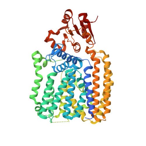
    Polymyxins are antibiotics used in the last line of defense to combat multidrug-resistant infections by Gram-negative bacteria. Polymyxin resistance arises through charge modification of the bacterial outer membrane with the attachment of the cationic sugar 4-amino-4-deoxy-l-arabinose to lipid A, a reaction catalyzed by the integral membrane lipid-to-lipid glycosyltransferase 4-amino-4-deoxy-L-arabinose transferase (ArnT). Here, we report crystal structures of ArnT from Cupriavidus metallidurans, alone and in complex with the lipid carrier undecaprenyl phosphate, at 2.8 and 3.2 angstrom resolution, respectively. The structures show cavities for both lipidic substrates, which converge at the active site. A structural rearrangement occurs on undecaprenyl phosphate binding, which stabilizes the active site and likely allows lipid A binding. Functional mutagenesis experiments based on these structures suggest a mechanistic model for ArnT family enzymes.


  • Organizational Affiliation

    Department of Physiology and Cellular Biophysics, Columbia University, New York, NY 10032, USA.


Macromolecules
Find similar proteins by:  (by identity cutoff)  |  3D Structure
Entity ID: 1
MoleculeChains Sequence LengthOrganismDetailsImage
4-amino-4-deoxy-L-arabinose transferase or related glycosyltransferases of PMT family578Cupriavidus metallidurans CH34Mutation(s): 0 
Gene Names: Rmet_4828
EC: 2.4.2.43
Membrane Entity: Yes 
UniProt
Find proteins for Q1LDT6 (Cupriavidus metallidurans (strain ATCC 43123 / DSM 2839 / NBRC 102507 / CH34))
Explore Q1LDT6 
Go to UniProtKB:  Q1LDT6
Entity Groups  
Sequence Clusters30% Identity50% Identity70% Identity90% Identity95% Identity100% Identity
UniProt GroupQ1LDT6
Sequence Annotations
Expand
  • Reference Sequence
Small Molecules
Ligands 7 Unique
IDChains Name / Formula / InChI Key2D Diagram3D Interactions
DSL
Query on DSL

Download Ideal Coordinates CCD File 
C [auth A]MONO-TRANS, OCTA-CIS DECAPRENYL-PHOSPHATE
C50 H83 O4 P
XBEJBEIXLWRYBT-DJNGBRKISA-N
MPG
Query on MPG

Download Ideal Coordinates CCD File 
AA [auth A]
BA [auth A]
CA [auth A]
D [auth A]
DA [auth A]
AA [auth A],
BA [auth A],
CA [auth A],
D [auth A],
DA [auth A],
E [auth A],
EA [auth A],
F [auth A],
FA [auth A],
G [auth A],
GA [auth A],
H [auth A],
HA [auth A],
I [auth A],
IA [auth A],
J [auth A],
JA [auth A],
K [auth A],
KA [auth A],
L [auth A],
LA [auth A],
M [auth A],
MA [auth A],
N [auth A],
NA [auth A],
O [auth A],
OA [auth A],
P [auth A],
PA [auth A],
Q [auth A],
QA [auth A],
R [auth A],
RA [auth A],
S [auth A],
SA [auth A],
T [auth A],
TA [auth A],
U [auth A],
UA [auth A],
V [auth A],
VA [auth A],
W [auth A],
WA [auth A],
X [auth A],
XA [auth A],
Y [auth A],
YA [auth A],
Z [auth A]
[(Z)-octadec-9-enyl] (2R)-2,3-bis(oxidanyl)propanoate
C21 H40 O4
JPJYKWFFJCWMPK-GDCKJWNLSA-N
EPE
Query on EPE

Download Ideal Coordinates CCD File 
EB [auth A]4-(2-HYDROXYETHYL)-1-PIPERAZINE ETHANESULFONIC ACID
C8 H18 N2 O4 S
JKMHFZQWWAIEOD-UHFFFAOYSA-N
PC
Query on PC

Download Ideal Coordinates CCD File 
DB [auth A]PHOSPHOCHOLINE
C5 H15 N O4 P
YHHSONZFOIEMCP-UHFFFAOYSA-O
PO4
Query on PO4

Download Ideal Coordinates CCD File 
BB [auth A],
CB [auth A]
PHOSPHATE ION
O4 P
NBIIXXVUZAFLBC-UHFFFAOYSA-K
ZN
Query on ZN

Download Ideal Coordinates CCD File 
B [auth A]ZINC ION
Zn
PTFCDOFLOPIGGS-UHFFFAOYSA-N
CL
Query on CL

Download Ideal Coordinates CCD File 
AB [auth A],
ZA [auth A]
CHLORIDE ION
Cl
VEXZGXHMUGYJMC-UHFFFAOYSA-M
Experimental Data & Validation

Experimental Data

  • Method: X-RAY DIFFRACTION
  • Resolution: 2.70 Å
  • R-Value Free: 0.260 
  • R-Value Work: 0.212 
  • R-Value Observed: 0.215 
  • Space Group: P 21 21 21
Unit Cell:
Length ( Å )Angle ( ˚ )
a = 58.604α = 90
b = 80.63β = 90
c = 150.253γ = 90
Software Package:
Software NamePurpose
Aimlessdata scaling
PHENIXrefinement
PDB_EXTRACTdata extraction
iMOSFLMdata reduction
PHASERphasing

Structure Validation

View Full Validation Report


Ligand Structure Quality Assessment 


Entry History & Funding Information

Deposition Data


Funding OrganizationLocationGrant Number
National Institutes of Health/National Institute of General Medical Sciences (NIH/NIGMS)United StatesU54 GM095315
National Institutes of Health/National Institute of General Medical Sciences (NIH/NIGMS)United StatesR01 GM111980

Revision History  (Full details and data files)

  • Version 1.0: 2016-02-17
    Type: Initial release
  • Version 1.1: 2017-09-13
    Changes: Author supporting evidence, Database references, Derived calculations
  • Version 1.2: 2018-04-18
    Changes: Data collection, Database references
  • Version 1.3: 2019-12-25
    Changes: Author supporting evidence
  • Version 1.4: 2024-03-06
    Changes: Data collection, Database references