5EUN

The Crystal Structure of Human Kynurenine Aminotransferase II, PLP-bound form, at 1.83 A


Experimental Data Snapshot

  • Method: X-RAY DIFFRACTION
  • Resolution: 1.82 Å
  • R-Value Free: 0.199 
  • R-Value Work: 0.173 
  • R-Value Observed: 0.174 

wwPDB Validation   3D Report Full Report


This is version 1.3 of the entry. See complete history


Literature

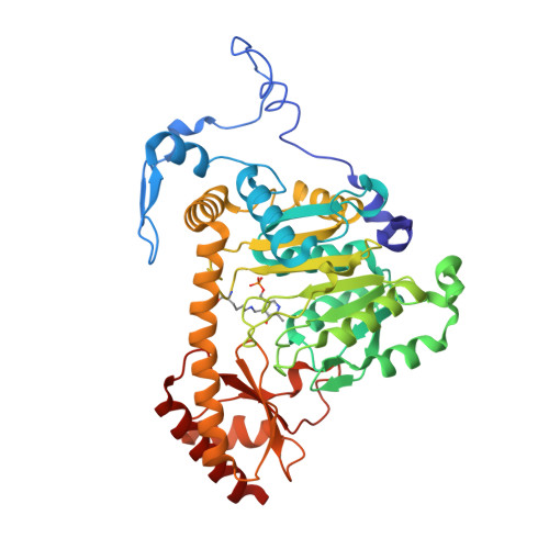
Structure of the PLP-Form of the Human Kynurenine Aminotransferase II in a Novel Spacegroup at 1.83 angstrom Resolution.

Nematollahi, A.Sun, G.Harrop, S.J.Hanrahan, J.R.Church, W.B.

(2016) Int J Mol Sci 17: 446-446

  • DOI: https://doi.org/10.3390/ijms17040446
  • Primary Citation of Related Structures:  
    5EUN

  • PubMed Abstract: 

    Kynurenine aminotransferase II (KAT-II) is a 47 kDa pyridoxal phosphate (PLP)-dependent enzyme, active as a homodimer, which catalyses the transamination of the amino acids kynurenine (KYN) and 3-hydroxykynurenine (3-HK) in the tryptophan pathway, and is responsible for producing metabolites that lead to kynurenic acid (KYNA), which is implicated in several neurological diseases such as schizophrenia. In order to fully describe the role of KAT-II in the pathobiology of schizophrenia and other brain disorders, the crystal structure of full-length PLP-form hKAT-II was determined at 1.83 Å resolution, the highest available. The electron density of the active site reveals an aldimine linkage between PLP and Lys263, as well as the active site residues, which characterize the fold-type I PLP-dependent enzymes.


  • Organizational Affiliation

    Group in Biomolecular Structure and Informatics, Faculty of Pharmacy, University of Sydney, Sydney, NSW 2006, Australia. anem7250@uni.sydney.edu.au.


Macromolecules
Find similar proteins by:  (by identity cutoff)  |  3D Structure
Entity ID: 1
MoleculeChains Sequence LengthOrganismDetailsImage
Kynurenine/alpha-aminoadipate aminotransferase, mitochondrial425Homo sapiensMutation(s): 0 
Gene Names: AADATKAT2
EC: 2.6.1.39 (PDB Primary Data), 2.6.1.7 (PDB Primary Data)
UniProt & NIH Common Fund Data Resources
Find proteins for Q8N5Z0 (Homo sapiens)
Explore Q8N5Z0 
Go to UniProtKB:  Q8N5Z0
PHAROS:  Q8N5Z0
GTEx:  ENSG00000109576 
Entity Groups  
Sequence Clusters30% Identity50% Identity70% Identity90% Identity95% Identity100% Identity
UniProt GroupQ8N5Z0
Sequence Annotations
Expand
  • Reference Sequence
Small Molecules
Modified Residues  1 Unique
IDChains TypeFormula2D DiagramParent
LLP
Query on LLP
A
L-PEPTIDE LINKINGC14 H22 N3 O7 PLYS
Experimental Data & Validation

Experimental Data

  • Method: X-RAY DIFFRACTION
  • Resolution: 1.82 Å
  • R-Value Free: 0.199 
  • R-Value Work: 0.173 
  • R-Value Observed: 0.174 
  • Space Group: P 43 21 2
Unit Cell:
Length ( Å )Angle ( ˚ )
a = 102.455α = 90
b = 102.455β = 90
c = 86.236γ = 90
Software Package:
Software NamePurpose
PHENIXrefinement
Blu-Icedata collection
XDSdata reduction
Aimlessdata scaling
PHENIXphasing

Structure Validation

View Full Validation Report


Entry History 

Deposition Data

Revision History  (Full details and data files)

  • Version 1.0: 2015-12-16
    Type: Initial release
  • Version 1.1: 2016-03-02
    Changes: Data collection
  • Version 1.2: 2016-04-13
    Changes: Database references
  • Version 1.3: 2016-08-10
    Changes: Database references