5ETO

E. coli 6-hydroxymethyl-7,8-dihydropterin pyrophosphokinase complexed with AMPCPP and inhibitor at 1.07 angstrom resolution


Experimental Data Snapshot

  • Method: X-RAY DIFFRACTION
  • Resolution: 1.07 Å
  • R-Value Free: 0.150 
  • R-Value Work: 0.129 
  • R-Value Observed: 0.130 

wwPDB Validation   3D Report Full Report


Ligand Structure Quality Assessment 


This is version 1.2 of the entry. See complete history


Literature

Structural Basis for the Selective Binding of Inhibitors to 6-Hydroxymethyl-7,8-dihydropterin Pyrophosphokinase from Staphylococcus aureus and Escherichia coli.

Dennis, M.L.Pitcher, N.P.Lee, M.D.DeBono, A.J.Wang, Z.C.Harjani, J.R.Rahmani, R.Cleary, B.Peat, T.S.Baell, J.B.Swarbrick, J.D.

(2016) J Med Chem 59: 5248-5263

  • DOI: https://doi.org/10.1021/acs.jmedchem.6b00002
  • Primary Citation of Related Structures:  
    5ETK, 5ETL, 5ETM, 5ETN, 5ETO, 5ETP, 5ETQ, 5ETR, 5ETS, 5ETT, 5ETV

  • PubMed Abstract: 

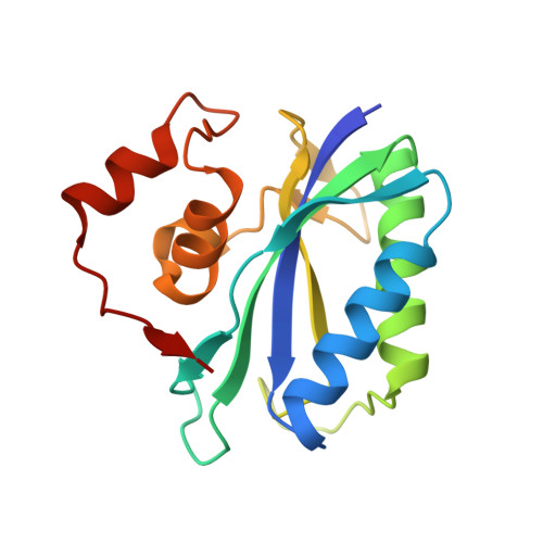
    6-Hydroxymethyl-7,8-dihydropterin pyrophosphokinase (HPPK) is a member of the folate biosynthesis pathway found in prokaryotes and lower eukaryotes that catalyzes the pyrophosphoryl transfer from the ATP cofactor to a 6-hydroxymethyl-7,8-dihydropterin substrate. We report the chemical synthesis of a series of S-functionalized 8-mercaptoguanine (8MG) analogues as substrate site inhibitors of HPPK and quantify binding against the E. coli and S. aureus enzymes (EcHPPK and SaHPPK). The results demonstrate that analogues incorporating acetophenone-based substituents have comparable affinities for both enzymes. Preferential binding of benzyl-substituted 8MG derivatives to SaHPPK was reconciled when a cryptic pocket unique to SaHPPK was revealed by X-ray crystallography. Differential chemical shift perturbation analysis confirmed this to be a common mode of binding for this series to SaHPPK. One compound (41) displayed binding affinities of 120 nM and 1.76 μM for SaHPPK and EcHPPK, respectively, and represents a lead for the development of more potent and selective inhibitors of SaHPPK.


  • Organizational Affiliation

    Monash Institute of Pharmaceutical Sciences, Monash University , Parkville, Victoria 3052, Australia.


Macromolecules
Find similar proteins by:  (by identity cutoff)  |  3D Structure
Entity ID: 1
MoleculeChains Sequence LengthOrganismDetailsImage
2-amino-4-hydroxy-6-hydroxymethyldihydropteridine pyrophosphokinase162Escherichia coliMutation(s): 0 
Gene Names: folKb0142JW0138
EC: 2.7.6.3
UniProt
Find proteins for P26281 (Escherichia coli (strain K12))
Explore P26281 
Go to UniProtKB:  P26281
Entity Groups  
Sequence Clusters30% Identity50% Identity70% Identity90% Identity95% Identity100% Identity
UniProt GroupP26281
Sequence Annotations
Expand
  • Reference Sequence
Small Molecules
Ligands 4 Unique
IDChains Name / Formula / InChI Key2D Diagram3D Interactions
APC
Query on APC

Download Ideal Coordinates CCD File 
C [auth A]DIPHOSPHOMETHYLPHOSPHONIC ACID ADENOSYL ESTER
C11 H18 N5 O12 P3
CAWZRIXWFRFUQB-IOSLPCCCSA-N
5RY
Query on 5RY

Download Ideal Coordinates CCD File 
B [auth A]4-[(2-azanyl-6-oxidanylidene-1,9-dihydropurin-8-yl)sulfanylmethyl]-3-fluoranyl-benzenecarbonitrile
C13 H9 F N6 O S
GEUVDHJEINQZML-UHFFFAOYSA-N
CA
Query on CA

Download Ideal Coordinates CCD File 
D [auth A],
E [auth A],
F [auth A],
G [auth A]
CALCIUM ION
Ca
BHPQYMZQTOCNFJ-UHFFFAOYSA-N
CL
Query on CL

Download Ideal Coordinates CCD File 
H [auth A],
I [auth A]
CHLORIDE ION
Cl
VEXZGXHMUGYJMC-UHFFFAOYSA-M
Binding Affinity Annotations 
IDSourceBinding Affinity
5RY BindingDB:  5ETO Kd: 2700 (nM) from 1 assay(s)
Binding MOAD:  5ETO Kd: 2700 (nM) from 1 assay(s)
Experimental Data & Validation

Experimental Data

  • Method: X-RAY DIFFRACTION
  • Resolution: 1.07 Å
  • R-Value Free: 0.150 
  • R-Value Work: 0.129 
  • R-Value Observed: 0.130 
  • Space Group: P 1 21 1
Unit Cell:
Length ( Å )Angle ( ˚ )
a = 35.77α = 90
b = 57.75β = 115.46
c = 38.42γ = 90
Software Package:
Software NamePurpose
REFMACrefinement
Cootmodel building
Aimlessdata scaling
MOLREPphasing
XDSdata reduction

Structure Validation

View Full Validation Report


Ligand Structure Quality Assessment 


Entry History 

Deposition Data

Revision History  (Full details and data files)

  • Version 1.0: 2016-05-04
    Type: Initial release
  • Version 1.1: 2016-06-22
    Changes: Database references
  • Version 1.2: 2023-09-27
    Changes: Data collection, Database references, Derived calculations, Refinement description