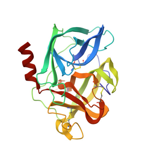
5EST

Crystallographic analysis of the inhibition of porcine pancreatic elastase by a peptidyl boronic acid: structure of a reaction intermediate


Experimental Data Snapshot

  • Method: X-RAY DIFFRACTION
  • Resolution: 2.09 Å
  • R-Value Work: 0.150 

wwPDB Validation   3D Report Full Report


Ligand Structure Quality Assessment 


This is version 1.3 of the entry. See complete history


Literature

Crystallographic analysis of the inhibition of porcine pancreatic elastase by a peptidyl boronic acid: structure of a reaction intermediate.

Takahashi, L.H.Radhakrishnan, R.Rosenfield Jr., R.E.Meyer Jr., E.F.

(1989) Biochemistry 28: 7610-7617

  • DOI: https://doi.org/10.1021/bi00445a016
  • Primary Citation of Related Structures:  
    5EST

  • PubMed Abstract: 

    The crystal structure of porcine pancreatic elastase (PPE) complexed to carbobenzoxy-alanylisoleucine-boronic acid (ZAIB) is reported to 2.09-A resolution and refined to an R factor of 0.15. This is the first reported structural analysis of PPE with an isoleucine residu in the primary specificity pocket. The results include (1) marked displacement of the inhibitor out of the active site leading to (2) a close (2.2 A) direct contact between B (boron atom of the inhibitor) and N epsilon of His-57 and also (3) covalent bonding (1.5 A) to O gamma of Ser-195. A scheme for the mechanism of inhibition of PPE by ZAIB is proposed. A comparison with a peptidyl difluoromethyl ketone-PPE complex (Ki = 9.5 microns) is made to explain the strong inhibition of PPE by ZAIB (Ki = 0.3 micron). These results lead us to characterize this structure as a time- and space-averaged reaction intermediate, providing fresh insight into the cramped dimensions available in enzymatic catalyses.


  • Organizational Affiliation

    Department of Chemistry, Texas A&M University, College Station 77843.


Macromolecules
Find similar proteins by:  (by identity cutoff)  |  3D Structure
Entity ID: 1
MoleculeChains Sequence LengthOrganismDetailsImage
ELASTASEA [auth E]240Sus scrofaMutation(s): 0 
EC: 3.4.21.36
UniProt
Find proteins for P00772 (Sus scrofa)
Explore P00772 
Go to UniProtKB:  P00772
Entity Groups  
Sequence Clusters30% Identity50% Identity70% Identity90% Identity95% Identity100% Identity
UniProt GroupP00772
Sequence Annotations
Expand
  • Reference Sequence
Small Molecules
Ligands 3 Unique
IDChains Name / Formula / InChI Key2D Diagram3D Interactions
0P2
Query on 0P2

Download Ideal Coordinates CCD File 
B [auth E]N~2~-[(benzyloxy)carbonyl]-N-[(1R,2S)-1-(dihydroxyboranyl)-2-methylbutyl]-L-alaninamide
C16 H25 B N2 O5
WMMGWXIYADLQHX-OBJOEFQTSA-N
SO4
Query on SO4

Download Ideal Coordinates CCD File 
C [auth E]SULFATE ION
O4 S
QAOWNCQODCNURD-UHFFFAOYSA-L
CA
Query on CA

Download Ideal Coordinates CCD File 
D [auth E]CALCIUM ION
Ca
BHPQYMZQTOCNFJ-UHFFFAOYSA-N
Experimental Data & Validation

Experimental Data

  • Method: X-RAY DIFFRACTION
  • Resolution: 2.09 Å
  • R-Value Work: 0.150 
  • Space Group: P 21 21 21
Unit Cell:
Length ( Å )Angle ( ˚ )
a = 52.28α = 90
b = 57.56β = 90
c = 75.41γ = 90
Software Package:
Software NamePurpose
EREFrefinement

Structure Validation

View Full Validation Report


Ligand Structure Quality Assessment 


Entry History 

Deposition Data

Revision History  (Full details and data files)

  • Version 1.0: 1992-04-15
    Type: Initial release
  • Version 1.1: 2008-03-03
    Changes: Version format compliance
  • Version 1.2: 2011-07-13
    Changes: Atomic model, Database references, Derived calculations, Non-polymer description, Structure summary, Version format compliance
  • Version 1.3: 2012-12-12
    Changes: Other