5ES1

CRYSTAL STRUCTURE OF MICROTUBULE AFFINITY-REGULATING KINASE 4 CATALYTIC DOMAIN IN COMPLEX WITH A PYRAZOLOPYRIMIDINE INHIBITOR


Experimental Data Snapshot

  • Method: X-RAY DIFFRACTION
  • Resolution: 2.80 Å
  • R-Value Free: 0.311 
  • R-Value Work: 0.239 
  • R-Value Observed: 0.243 

wwPDB Validation   3D Report Full Report


Ligand Structure Quality Assessment 


This is version 1.4 of the entry. See complete history


Literature

Crystal structure of microtubule affinity-regulating kinase 4 catalytic domain in complex with a pyrazolopyrimidine inhibitor.

Sack, J.S.Gao, M.Kiefer, S.E.Myers, J.E.Newitt, J.A.Wu, S.Yan, C.

(2016) Acta Crystallogr F Struct Biol Commun 72: 129-134

  • DOI: https://doi.org/10.1107/S2053230X15024747
  • Primary Citation of Related Structures:  
    5ES1

  • PubMed Abstract: 

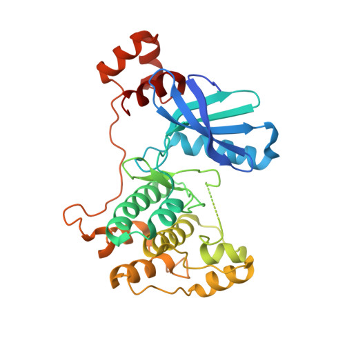
    Microtubule-associated protein/microtubule affinity-regulating kinase 4 (MARK4) is a serine/threonine kinase involved in the phosphorylation of MAP proteins that regulate microtubule dynamics. Abnormal activity of MARK4 has been proposed to contribute to neurofibrillary tangle formation in Alzheimer's disease. The crystal structure of the catalytic and ubiquitin-associated domains of MARK4 with a potent pyrazolopyrimidine inhibitor has been determined to 2.8 Å resolution with an Rwork of 22.8%. The overall structure of MARK4 is similar to those of the other known MARK isoforms. The inhibitor is located in the ATP-binding site, with the pyrazolopyrimidine group interacting with the inter-lobe hinge region while the aminocyclohexane moiety interacts with the catalytic loop and the DFG motif, forcing the activation loop out of the ATP-binding pocket.


  • Organizational Affiliation

    Molecular Discovery Technologies, Bristol-Myers Squibb Research and Development, PO Box 4000, Princeton, NJ 08543-4000, USA.


Macromolecules
Find similar proteins by:  (by identity cutoff)  |  3D Structure
Entity ID: 1
MoleculeChains Sequence LengthOrganismDetailsImage
MAP/microtubule affinity-regulating kinase 4328Homo sapiensMutation(s): 0 
Gene Names: MARK4KIAA1860MARKL1
EC: 2.7.11.1
UniProt & NIH Common Fund Data Resources
Find proteins for Q96L34 (Homo sapiens)
Explore Q96L34 
Go to UniProtKB:  Q96L34
PHAROS:  Q96L34
GTEx:  ENSG00000007047 
Entity Groups  
Sequence Clusters30% Identity50% Identity70% Identity90% Identity95% Identity100% Identity
UniProt GroupQ96L34
Sequence Annotations
Expand
  • Reference Sequence
Small Molecules
Ligands 1 Unique
IDChains Name / Formula / InChI Key2D Diagram3D Interactions
5RC
Query on 5RC

Download Ideal Coordinates CCD File 
B [auth A]~{N}-[(1~{R},6~{R})-6-azanyl-2,2-bis(fluoranyl)cyclohexyl]-5-ethyl-4-[6-(trifluoromethyl)pyrazolo[1,5-a]pyrimidin-3-yl]thiophene-2-carboxamide
C20 H20 F5 N5 O S
QXXZIVGTJXYLQO-CZUORRHYSA-N
Experimental Data & Validation

Experimental Data

  • Method: X-RAY DIFFRACTION
  • Resolution: 2.80 Å
  • R-Value Free: 0.311 
  • R-Value Work: 0.239 
  • R-Value Observed: 0.243 
  • Space Group: P 62
Unit Cell:
Length ( Å )Angle ( ˚ )
a = 111.478α = 90
b = 111.478β = 90
c = 69.775γ = 120
Software Package:
Software NamePurpose
DENZOdata reduction
BUSTER-TNTrefinement
PDB_EXTRACTdata extraction
SCALEPACKdata scaling
PHASERphasing

Structure Validation

View Full Validation Report


Ligand Structure Quality Assessment 


Entry History 

Deposition Data

  • Released Date: 2015-12-02 
  • Deposition Author(s): Sack, J.S.

Revision History  (Full details and data files)

  • Version 1.0: 2015-12-02
    Type: Initial release
  • Version 1.1: 2016-02-10
    Changes: Database references
  • Version 1.2: 2017-11-22
    Changes: Derived calculations, Refinement description
  • Version 1.3: 2018-04-18
    Changes: Data collection, Database references
  • Version 1.4: 2023-09-27
    Changes: Data collection, Database references, Refinement description