5EPO

The three-dimensional structure of Clostridium absonum 7alpha-hydroxysteroid dehydrogenase


Experimental Data Snapshot

  • Method: X-RAY DIFFRACTION
  • Resolution: 2.00 Å
  • R-Value Free: 0.222 
  • R-Value Work: 0.182 
  • R-Value Observed: 0.184 

wwPDB Validation   3D Report Full Report


Ligand Structure Quality Assessment 


This is version 1.2 of the entry. See complete history


Literature

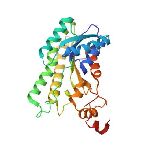
The three-dimensional structure of Clostridium absonum 7 alpha-hydroxysteroid dehydrogenase: new insights into the conserved arginines for NADP(H) recognition

Lou, D.Wang, B.Tan, J.Zhu, L.Cen, X.Ji, Q.Wang, Y.

(2016) Sci Rep 6: 22885-22885

  • DOI: https://doi.org/10.1038/srep22885
  • Primary Citation of Related Structures:  
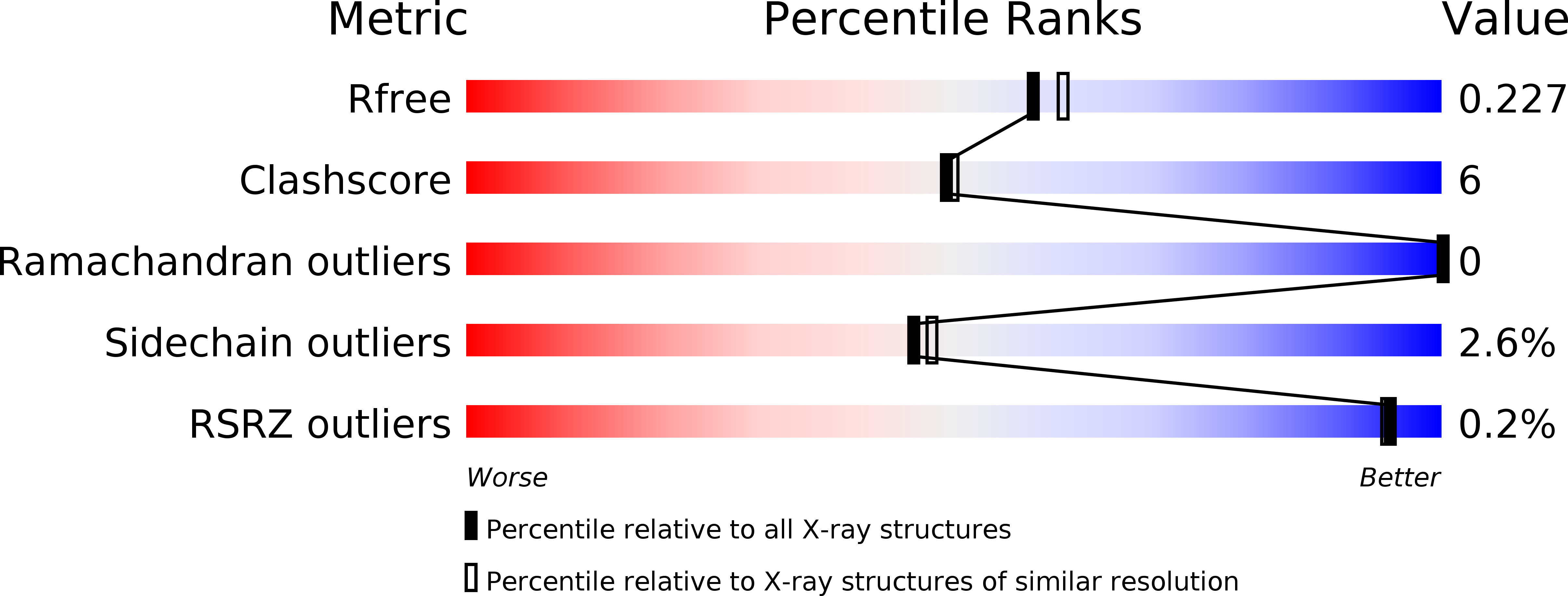
    5EPO

  • PubMed Abstract: 

    7α-hydroxysteroid dehydrogenase (7α-HSDH) can catalyse the oxidation of C7 α-OH of the steroid nucleus in the bile acid metabolism. In the paper we determined the crystal structure of 7α-HSDH from Clostridium absonum (CA 7α-HSDH) complexed with taurochenodeoxycholic acid (TCDCA) and NADP(+) by X-ray diffraction, which, as a tetramer, possesses the typical α/β folding pattern. The four subunits of an asymmetric unit lie in the fact that there are the stable hydrophobic interactions between Q-axis-related subunits. Significantly, we captured an active state of the NADP(+), confirming that nicotinamide moiety of NADP(+) act as electron carrier in the dehydrogenation. On the basis of crystal structure analysis, site-directed mutagenesis and MD simulation, furthermore, we find that the guanidinium of Arg38 can form the stable cation-π interaction with the adenine ring of NADP(+), and the cation-π interaction and hydrogen bonds between Arg38 and NADP(+) have a significant anchor effect on the cofactor binding to CA 7α-HSDH.


  • Organizational Affiliation

    Key Laboratory of Biorheological Science and Technology (Chongqing University), Ministry of Education, College of Bioengineering, Chongqing University, Chongqing 400030, China.


Macromolecules
Find similar proteins by:  (by identity cutoff)  |  3D Structure
Entity ID: 1
MoleculeChains Sequence LengthOrganismDetailsImage
7-alpha-hydroxysteroid deydrogenase
A, B, C, D
262Clostridium sardinienseMutation(s): 0 
UniProt
Find proteins for G9FRD7 (Clostridium sardiniense)
Explore G9FRD7 
Go to UniProtKB:  G9FRD7
Entity Groups  
Sequence Clusters30% Identity50% Identity70% Identity90% Identity95% Identity100% Identity
UniProt GroupG9FRD7
Sequence Annotations
Expand
  • Reference Sequence
Small Molecules
Ligands 3 Unique
IDChains Name / Formula / InChI Key2D Diagram3D Interactions
NAP
Query on NAP

Download Ideal Coordinates CCD File 
E [auth A],
H [auth B],
K [auth C],
N [auth D]
NADP NICOTINAMIDE-ADENINE-DINUCLEOTIDE PHOSPHATE
C21 H28 N7 O17 P3
XJLXINKUBYWONI-NNYOXOHSSA-N
TUD
Query on TUD

Download Ideal Coordinates CCD File 
F [auth A],
I [auth B],
L [auth C],
O [auth D]
TAUROCHENODEOXYCHOLIC ACID
C26 H45 N O6 S
BHTRKEVKTKCXOH-BJLOMENOSA-N
GOL
Query on GOL

Download Ideal Coordinates CCD File 
G [auth A],
J [auth B],
M [auth C],
P [auth D]
GLYCEROL
C3 H8 O3
PEDCQBHIVMGVHV-UHFFFAOYSA-N
Experimental Data & Validation

Experimental Data

  • Method: X-RAY DIFFRACTION
  • Resolution: 2.00 Å
  • R-Value Free: 0.222 
  • R-Value Work: 0.182 
  • R-Value Observed: 0.184 
  • Space Group: P 21 21 21
Unit Cell:
Length ( Å )Angle ( ˚ )
a = 67.18α = 90
b = 69.68β = 90
c = 220.39γ = 90
Software Package:
Software NamePurpose
REFMACrefinement
SCALAdata scaling
PHASERphasing

Structure Validation

View Full Validation Report


Ligand Structure Quality Assessment 


Entry History & Funding Information

Deposition Data


Funding OrganizationLocationGrant Number
China2014ZX09301306-007

Revision History  (Full details and data files)

  • Version 1.0: 2016-03-23
    Type: Initial release
  • Version 1.1: 2017-09-27
    Changes: Data collection, Derived calculations
  • Version 1.2: 2024-03-20
    Changes: Data collection, Database references