5EOF

Crystal structure of OPTN NTD and TBK1 CTD complex


Experimental Data Snapshot

  • Method: X-RAY DIFFRACTION
  • Resolution: 2.05 Å
  • R-Value Free: 0.245 
  • R-Value Work: 0.207 
  • R-Value Observed: 0.209 

wwPDB Validation   3D Report Full Report


This is version 1.1 of the entry. See complete history


Literature

Structural insights into the interaction and disease mechanism of neurodegenerative disease-associated optineurin and TBK1 proteins.

Li, F.Xie, X.Wang, Y.Liu, J.Cheng, X.Guo, Y.Gong, Y.Hu, S.Pan, L.

(2016) Nat Commun 7: 12708-12708

  • DOI: https://doi.org/10.1038/ncomms12708
  • Primary Citation of Related Structures:  
    5EOA, 5EOF, 5EP6

  • PubMed Abstract: 

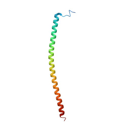
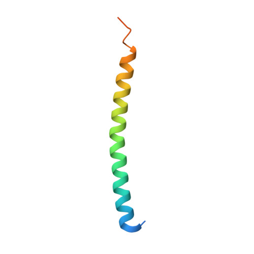
    Optineurin is an important autophagy receptor involved in several selective autophagy processes, during which its function is regulated by TBK1. Mutations of optineurin and TBK1 are both associated with neurodegenerative diseases. However, the mechanistic basis underlying the specific interaction between optineurin and TBK1 is still elusive. Here we determine the crystal structures of optineurin/TBK1 complex and the related NAP1/TBK1 complex, uncovering the detailed molecular mechanism governing the optineurin and TBK1 interaction, and revealing a general binding mode between TBK1 and its associated adaptor proteins. In addition, we demonstrate that the glaucoma-associated optineurin E50K mutation not only enhances the interaction between optineurin and TBK1 but also alters the oligomeric state of optineurin, and the ALS-related TBK1 E696K mutation specifically disrupts the optineurin/TBK1 complex formation but has little effect on the NAP1/TBK1 complex. Thus, our study provides mechanistic insights into those currently known disease-causing optineurin and TBK1 mutations found in patients.


  • Organizational Affiliation

    State Key Laboratory of Bioorganic and Natural Products Chemistry, Shanghai Institute of Organic Chemistry, Chinese Academy of Sciences, 345 Lingling Road, Shanghai 200032, China.


Macromolecules
Find similar proteins by:  (by identity cutoff)  |  3D Structure
Entity ID: 1
MoleculeChains Sequence LengthOrganismDetailsImage
Optineurin
A, B
82Homo sapiensMutation(s): 0 
Gene Names: OPTNFIP2GLC1EHIP7HYPLNRP
UniProt & NIH Common Fund Data Resources
Find proteins for Q96CV9 (Homo sapiens)
Explore Q96CV9 
Go to UniProtKB:  Q96CV9
PHAROS:  Q96CV9
GTEx:  ENSG00000123240 
Entity Groups  
Sequence Clusters30% Identity50% Identity70% Identity90% Identity95% Identity100% Identity
UniProt GroupQ96CV9
Sequence Annotations
Expand
  • Reference Sequence
Find similar proteins by:  (by identity cutoff)  |  3D Structure
Entity ID: 2
MoleculeChains Sequence LengthOrganismDetailsImage
Serine/threonine-protein kinase TBK1
C, D
57Homo sapiensMutation(s): 0 
Gene Names: TBK1NAK
EC: 2.7.11.1
UniProt & NIH Common Fund Data Resources
Find proteins for Q9UHD2 (Homo sapiens)
Explore Q9UHD2 
Go to UniProtKB:  Q9UHD2
PHAROS:  Q9UHD2
GTEx:  ENSG00000183735 
Entity Groups  
Sequence Clusters30% Identity50% Identity70% Identity90% Identity95% Identity100% Identity
UniProt GroupQ9UHD2
Sequence Annotations
Expand
  • Reference Sequence
Experimental Data & Validation

Experimental Data

  • Method: X-RAY DIFFRACTION
  • Resolution: 2.05 Å
  • R-Value Free: 0.245 
  • R-Value Work: 0.207 
  • R-Value Observed: 0.209 
  • Space Group: P 21 21 21
Unit Cell:
Length ( Å )Angle ( ˚ )
a = 36.321α = 90
b = 54.775β = 90
c = 153.141γ = 90
Software Package:
Software NamePurpose
PHENIXrefinement
HKL-2000data reduction
HKL-2000data scaling
PHENIXphasing

Structure Validation

View Full Validation Report


Entry History & Funding Information

Deposition Data


Funding OrganizationLocationGrant Number
National Natural Science Foundation of ChinaChina31470749
National Basic Research Program of ChinaChina2013CB836900

Revision History  (Full details and data files)

  • Version 1.0: 2016-09-28
    Type: Initial release
  • Version 1.1: 2024-03-20
    Changes: Data collection, Database references, Derived calculations