5EN2

Molecular basis for antibody-mediated neutralization of New World hemorrhagic fever mammarenaviruses


Experimental Data Snapshot

  • Method: X-RAY DIFFRACTION
  • Resolution: 1.82 Å
  • R-Value Free: 0.224 
  • R-Value Work: 0.180 
  • R-Value Observed: 0.183 

wwPDB Validation   3D Report Full Report


Ligand Structure Quality Assessment 


This is version 2.1 of the entry. See complete history


Literature

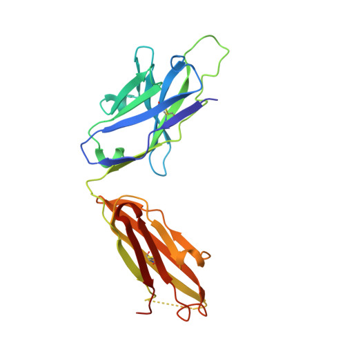
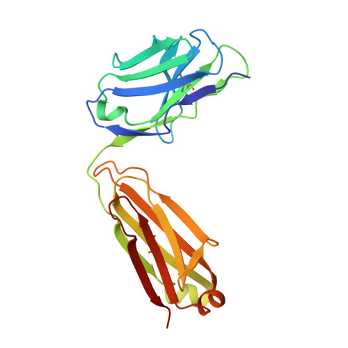
Molecular Basis for Antibody-Mediated Neutralization of New World Hemorrhagic Fever Mammarenaviruses.

Mahmutovic, S.Clark, L.Levis, S.C.Briggiler, A.M.Enria, D.A.Harrison, S.C.Abraham, J.

(2015) Cell Host Microbe 18: 705-713

  • DOI: https://doi.org/10.1016/j.chom.2015.11.005
  • Primary Citation of Related Structures:  
    5EN2

  • PubMed Abstract: 

    In the Western hemisphere, at least five mammarenaviruses cause human viral hemorrhagic fevers with high case fatality rates. Junín virus (JUNV) is the only hemorrhagic fever virus for which transfusion of survivor immune plasma that contains neutralizing antibodies ("passive immunity") is an established treatment. Here, we report the structure of the JUNV surface glycoprotein receptor-binding subunit (GP1) bound to a neutralizing monoclonal antibody. The antibody engages the GP1 site that binds transferrin receptor 1 (TfR1)-the host cell surface receptor for all New World hemorrhagic fever mammarenaviruses-and mimics an important receptor contact. We show that survivor immune plasma contains antibodies that bind the same epitope. We propose that viral receptor-binding site accessibility explains the success of passive immunity against JUNV and that this functionally conserved epitope is a potential target for therapeutics and vaccines to limit infection by all New World hemorrhagic fever mammarenaviruses.


  • Organizational Affiliation

    Laboratory of Molecular Medicine, Boston Children's Hospital, Harvard Medical School, Boston, MA 02115, USA.


Macromolecules
Find similar proteins by:  (by identity cutoff)  |  3D Structure
Entity ID: 1
MoleculeChains Sequence LengthOrganismDetailsImage
GD01 heavy chain224Mus musculusMutation(s): 0 
Entity Groups  
Sequence Clusters30% Identity50% Identity70% Identity90% Identity95% Identity100% Identity
Sequence Annotations
Expand
  • Reference Sequence
Find similar proteins by:  (by identity cutoff)  |  3D Structure
Entity ID: 2
MoleculeChains Sequence LengthOrganismDetailsImage
GD01 light chain212Mus musculusMutation(s): 0 
Entity Groups  
Sequence Clusters30% Identity50% Identity70% Identity90% Identity95% Identity100% Identity
Sequence Annotations
Expand
  • Reference Sequence
Find similar proteins by:  (by identity cutoff)  |  3D Structure
Entity ID: 3
MoleculeChains Sequence LengthOrganismDetailsImage
Pre-glycoprotein polyprotein GP complex141Mammarenavirus juninenseMutation(s): 0 
Gene Names: GPCGP-C
UniProt
Find proteins for P26313 (Junin mammarenavirus)
Explore P26313 
Go to UniProtKB:  P26313
Entity Groups  
Sequence Clusters30% Identity50% Identity70% Identity90% Identity95% Identity100% Identity
UniProt GroupP26313
Sequence Annotations
Expand
  • Reference Sequence
Oligosaccharides

Help

Entity ID: 4
MoleculeChains Length2D Diagram Glycosylation3D Interactions
alpha-D-mannopyranose-(1-3)-[alpha-D-mannopyranose-(1-6)]alpha-D-mannopyranose-(1-6)-[alpha-D-mannopyranose-(1-3)]beta-D-mannopyranose-(1-4)-2-acetamido-2-deoxy-beta-D-glucopyranose-(1-4)-2-acetamido-2-deoxy-beta-D-glucopyranose
D
7N-Glycosylation
Glycosylation Resources
GlyTouCan:  G55220VL
GlyCosmos:  G55220VL
GlyGen:  G55220VL
Entity ID: 5
MoleculeChains Length2D Diagram Glycosylation3D Interactions
2-acetamido-2-deoxy-beta-D-glucopyranose-(1-4)-2-acetamido-2-deoxy-beta-D-glucopyranose
E
2N-Glycosylation
Glycosylation Resources
GlyTouCan:  G42666HT
GlyCosmos:  G42666HT
GlyGen:  G42666HT
Small Molecules
Ligands 2 Unique
IDChains Name / Formula / InChI Key2D Diagram3D Interactions
NAG
Query on NAG

Download Ideal Coordinates CCD File 
G [auth C]2-acetamido-2-deoxy-beta-D-glucopyranose
C8 H15 N O6
OVRNDRQMDRJTHS-FMDGEEDCSA-N
CL
Query on CL

Download Ideal Coordinates CCD File 
F [auth B]CHLORIDE ION
Cl
VEXZGXHMUGYJMC-UHFFFAOYSA-M
Experimental Data & Validation

Experimental Data

  • Method: X-RAY DIFFRACTION
  • Resolution: 1.82 Å
  • R-Value Free: 0.224 
  • R-Value Work: 0.180 
  • R-Value Observed: 0.183 
  • Space Group: P 21 21 21
Unit Cell:
Length ( Å )Angle ( ˚ )
a = 52.11α = 90
b = 74.79β = 90
c = 177.64γ = 90
Software Package:
Software NamePurpose
BUSTER-TNTrefinement
iMOSFLMdata reduction
SCALAdata scaling
PHASERphasing
PDB_EXTRACTdata extraction

Structure Validation

View Full Validation Report


Ligand Structure Quality Assessment 


Entry History & Funding Information

Deposition Data


Funding OrganizationLocationGrant Number
National Institutes of Health/National Institute Of Allergy and Infectious Diseases (NIH/NIAID)United StatesAI-109740

Revision History  (Full details and data files)

  • Version 1.0: 2015-12-30
    Type: Initial release
  • Version 1.1: 2017-09-13
    Changes: Author supporting evidence, Derived calculations
  • Version 1.2: 2019-12-11
    Changes: Author supporting evidence
  • Version 2.0: 2020-07-29
    Type: Remediation
    Reason: Carbohydrate remediation
    Changes: Advisory, Atomic model, Data collection, Derived calculations, Structure summary
  • Version 2.1: 2023-09-27
    Changes: Data collection, Database references, Derived calculations, Refinement description, Structure summary