5ELV

Crystal structure of the GluA2 ligand-binding domain (S1S2J-L504-N775) in complex with glutamate and BPAM-521 at 1.92 A resolution


Experimental Data Snapshot

  • Method: X-RAY DIFFRACTION
  • Resolution: 1.92 Å
  • R-Value Free: 0.209 
  • R-Value Work: 0.162 
  • R-Value Observed: 0.164 

wwPDB Validation   3D Report Full Report


Ligand Structure Quality Assessment 


This is version 1.4 of the entry. See complete history


Literature

Enthalpy-Entropy Compensation in the Binding of Modulators at Ionotropic Glutamate Receptor GluA2.

Krintel, C.Francotte, P.Pickering, D.S.Juknaite, L.Phlsgaard, J.Olsen, L.Frydenvang, K.Goffin, E.Pirotte, B.Kastrup, J.S.

(2016) Biophys J 110: 2397-2406

  • DOI: https://doi.org/10.1016/j.bpj.2016.04.032
  • Primary Citation of Related Structures:  
    5ELV

  • PubMed Abstract: 

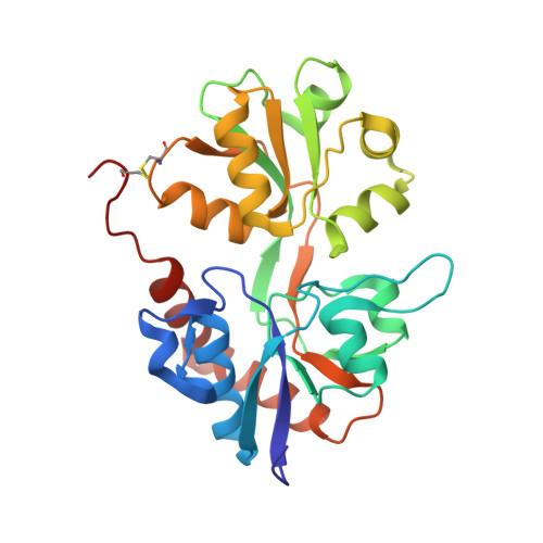
    The 1,2,4-benzothiadiazine 1,1-dioxide type of positive allosteric modulators of the ionotropic glutamate receptor A2 (GluA2) are promising lead compounds for the treatment of cognitive disorders, e.g., Alzheimer's disease. The modulators bind in a cleft formed by the interface of two neighboring ligand binding domains and act by stabilizing the agonist-bound open-channel conformation. The driving forces behind the binding of these modulators can be significantly altered with only minor substitutions to the parent molecules. In this study, we show that changing the 7-fluorine substituent of modulators BPAM97 (2) and BPAM344 (3) into a hydroxyl group (BPAM557 (4) and BPAM521 (5), respectively), leads to a more favorable binding enthalpy (ΔH, kcal/mol) from -4.9 (2) and -7.5 (3) to -6.2 (4) and -14.5 (5), but also a less favorable binding entropy (-TΔS, kcal/mol) from -2.3 (2) and -1.3 (3) to -0.5 (4) and 4.8 (5). Thus, the dissociation constants (Kd, μM) of 4 (11.2) and 5 (0.16) are similar to those of 2 (5.6) and 3 (0.35). Functionally, 4 and 5 potentiated responses of 10 μM L-glutamate at homomeric rat GluA2(Q)i receptors with EC50 values of 67.3 and 2.45 μM, respectively. The binding mode of 5 was examined with x-ray crystallography, showing that the only change compared to that of earlier compounds was the orientation of Ser-497 pointing toward the hydroxyl group of 5. The favorable enthalpy can be explained by the formation of a hydrogen bond from the side-chain hydroxyl group of Ser-497 to the hydroxyl group of 5, whereas the unfavorable entropy might be due to desolvation effects combined with a conformational restriction of Ser-497 and 5. In summary, this study shows a remarkable example of enthalpy-entropy compensation in drug development accompanied with a likely explanation of the underlying structural mechanism.


  • Organizational Affiliation

    Department of Drug Design and Pharmacology, Faculty of Health and Medical Sciences, University of Copenhagen, Copenhagen, Denmark.


Macromolecules
Find similar proteins by:  (by identity cutoff)  |  3D Structure
Entity ID: 1
MoleculeChains Sequence LengthOrganismDetailsImage
Glutamate receptor 2,Glutamate receptor 2
A, B
264Rattus norvegicusMutation(s): 2 
Gene Names: Gria2Glur2
UniProt
Find proteins for P19491 (Rattus norvegicus)
Explore P19491 
Go to UniProtKB:  P19491
Entity Groups  
Sequence Clusters30% Identity50% Identity70% Identity90% Identity95% Identity100% Identity
UniProt GroupP19491
Sequence Annotations
Expand
  • Reference Sequence
Small Molecules
Ligands 7 Unique
IDChains Name / Formula / InChI Key2D Diagram3D Interactions
5PX
Query on 5PX

Download Ideal Coordinates CCD File 
AA [auth B],
N [auth A]
4-Cyclopropyl-3,4-dihydro-7-hydroxy-2H-1,2,4-benzothiadiazine 1,1-dioxide
C10 H12 N2 O3 S
OMEAYSCNDLQLNC-UHFFFAOYSA-N
GLU
Query on GLU

Download Ideal Coordinates CCD File 
BA [auth B],
O [auth A]
GLUTAMIC ACID
C5 H9 N O4
WHUUTDBJXJRKMK-VKHMYHEASA-N
PEG
Query on PEG

Download Ideal Coordinates CCD File 
Y [auth B],
Z [auth B]
DI(HYDROXYETHYL)ETHER
C4 H10 O3
MTHSVFCYNBDYFN-UHFFFAOYSA-N
SO4
Query on SO4

Download Ideal Coordinates CCD File 
C [auth A],
D [auth A],
E [auth A],
P [auth B]
SULFATE ION
O4 S
QAOWNCQODCNURD-UHFFFAOYSA-L
GOL
Query on GOL

Download Ideal Coordinates CCD File 
F [auth A],
G [auth A],
Q [auth B],
R [auth B]
GLYCEROL
C3 H8 O3
PEDCQBHIVMGVHV-UHFFFAOYSA-N
ACT
Query on ACT

Download Ideal Coordinates CCD File 
J [auth A]
K [auth A]
L [auth A]
M [auth A]
T [auth B]
J [auth A],
K [auth A],
L [auth A],
M [auth A],
T [auth B],
U [auth B],
V [auth B],
W [auth B],
X [auth B]
ACETATE ION
C2 H3 O2
QTBSBXVTEAMEQO-UHFFFAOYSA-M
CL
Query on CL

Download Ideal Coordinates CCD File 
H [auth A],
I [auth A],
S [auth B]
CHLORIDE ION
Cl
VEXZGXHMUGYJMC-UHFFFAOYSA-M
Binding Affinity Annotations 
IDSourceBinding Affinity
5PX Binding MOAD:  5ELV Kd: 160 (nM) from 1 assay(s)
Experimental Data & Validation

Experimental Data

  • Method: X-RAY DIFFRACTION
  • Resolution: 1.92 Å
  • R-Value Free: 0.209 
  • R-Value Work: 0.162 
  • R-Value Observed: 0.164 
  • Space Group: P 21 21 2
Unit Cell:
Length ( Å )Angle ( ˚ )
a = 98.79α = 90
b = 122.48β = 90
c = 47.54γ = 90
Software Package:
Software NamePurpose
SCALAdata scaling
PHASERphasing
PHENIXrefinement
PDB_EXTRACTdata extraction
XDSdata reduction
Cootmodel building

Structure Validation

View Full Validation Report


Ligand Structure Quality Assessment 


Entry History 

Deposition Data

Revision History  (Full details and data files)

  • Version 1.0: 2016-05-04
    Type: Initial release
  • Version 1.1: 2016-06-22
    Changes: Database references
  • Version 1.2: 2018-01-17
    Changes: Data collection
  • Version 1.3: 2018-03-07
    Changes: Source and taxonomy
  • Version 1.4: 2024-01-10
    Changes: Data collection, Database references, Refinement description