5EKO

Crystal structure of MAPK13 complex with inhibitor


Experimental Data Snapshot

  • Method: X-RAY DIFFRACTION
  • Resolution: 2.00 Å
  • R-Value Free: 0.241 
  • R-Value Work: 0.200 
  • R-Value Observed: 0.202 

wwPDB Validation   3D Report Full Report


Ligand Structure Quality Assessment 


This is version 1.6 of the entry. See complete history


Literature

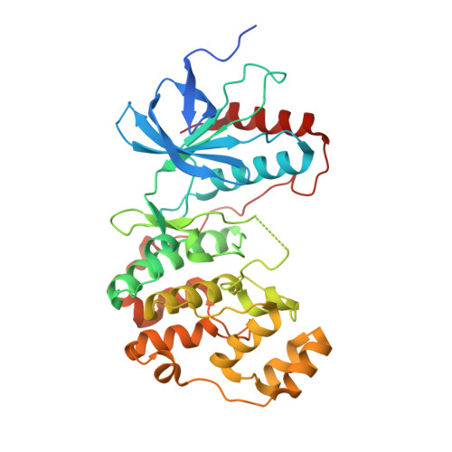
First comprehensive structural and biophysical analysis of MAPK13 inhibitors targeting DFG-in and DFG-out binding modes.

Yurtsever, Z.Patel, D.A.Kober, D.L.Su, A.Miller, C.A.Romero, A.G.Holtzman, M.J.Brett, T.J.

(2016) Biochim Biophys Acta 1860: 2335-2344

  • DOI: https://doi.org/10.1016/j.bbagen.2016.06.023
  • Primary Citation of Related Structures:  
    5EKN, 5EKO

  • PubMed Abstract: 

    P38 MAP kinases are centrally involved in mediating extracellular signaling in various diseases. While much attention has previously been focused on the ubiquitously expressed family member MAPK14 (p38α), recent studies indicate that family members such as MAPK13 (p38δ) display a more selective cellular and tissue expression and might therefore represent a specific kinase to target in certain diseases.


  • Organizational Affiliation

    Division of Pulmonary and Critical Care Medicine, Department of Medicine, Washington University School of Medicine, St. Louis, MO 63110, United States; Biochemistry Program, Washington University School of Medicine, St. Louis, MO 63110, United States; Center for the Investigation of Membrane Excitability Diseases, Washington University School of Medicine, St. Louis, MO 63110, United States.


Macromolecules
Find similar proteins by:  (by identity cutoff)  |  3D Structure
Entity ID: 1
MoleculeChains Sequence LengthOrganismDetailsImage
Mitogen-activated protein kinase 13371Homo sapiensMutation(s): 0 
Gene Names: MAPK13PRKM13SAPK4
EC: 2.7.11.24
UniProt & NIH Common Fund Data Resources
Find proteins for O15264 (Homo sapiens)
Explore O15264 
Go to UniProtKB:  O15264
PHAROS:  O15264
GTEx:  ENSG00000156711 
Entity Groups  
Sequence Clusters30% Identity50% Identity70% Identity90% Identity95% Identity100% Identity
UniProt GroupO15264
Sequence Annotations
Expand
  • Reference Sequence
Small Molecules
Ligands 1 Unique
IDChains Name / Formula / InChI Key2D Diagram3D Interactions
N17
Query on N17

Download Ideal Coordinates CCD File 
B [auth A]3-(4-methyl-1H-imidazol-1-yl)-N-[4-(pyridin-4-yloxy)phenyl]benzamide
C22 H18 N4 O2
TXKSFDQJSFSHRB-UHFFFAOYSA-N
Binding Affinity Annotations 
IDSourceBinding Affinity
N17 BindingDB:  5EKO IC50: min: 1.02e+4, max: 1.58e+4 (nM) from 2 assay(s)
Binding MOAD:  5EKO IC50: 1.58e+4 (nM) from 1 assay(s)
Experimental Data & Validation

Experimental Data

Unit Cell:
Length ( Å )Angle ( ˚ )
a = 61.263α = 90
b = 69.669β = 90
c = 93.068γ = 90
Software Package:
Software NamePurpose
PHENIXrefinement
HKL-2000data reduction
SCALEPACKdata scaling
PHASERphasing

Structure Validation

View Full Validation Report


Ligand Structure Quality Assessment 


Entry History & Funding Information

Deposition Data


Funding OrganizationLocationGrant Number
National Institutes of Health/National Heart, Lung, and Blood Institute (NIH/NHLBI)United StatesR01-HL119813

Revision History  (Full details and data files)

  • Version 1.0: 2016-07-06
    Type: Initial release
  • Version 1.1: 2016-07-20
    Changes: Database references
  • Version 1.2: 2016-09-14
    Changes: Database references
  • Version 1.3: 2017-09-20
    Changes: Author supporting evidence, Database references, Derived calculations
  • Version 1.4: 2017-11-01
    Changes: Author supporting evidence
  • Version 1.5: 2019-12-04
    Changes: Author supporting evidence
  • Version 1.6: 2023-09-27
    Changes: Data collection, Database references, Refinement description