


structure of the autoinhibited Epha2 JMS-KD
Wei, Q., Zheng, J.To be published.
Experimental Data Snapshot
wwPDB Validation 3D Report Full Report

Entity ID: 1 | |||||
|---|---|---|---|---|---|
| Molecule | Chains | Sequence Length | Organism | Details | Image |
| Ephrin type-A receptor 2 | 308 | Homo sapiens | Mutation(s): 0 Gene Names: EPHA2, ECK EC: 2.7.10.1 | 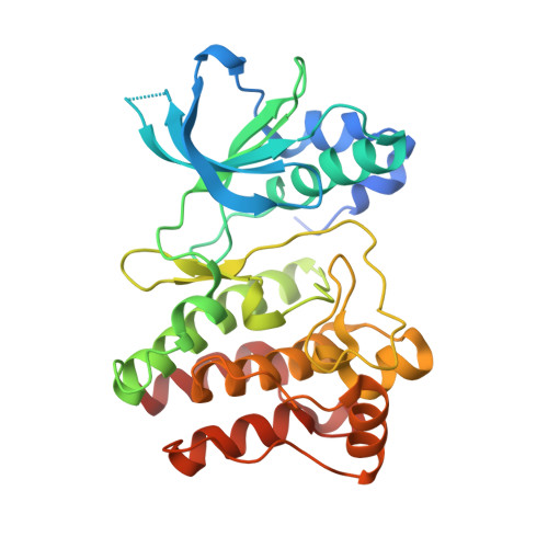 | |
UniProt & NIH Common Fund Data Resources | |||||
Find proteins for P29317 (Homo sapiens) Explore P29317 Go to UniProtKB: P29317 | |||||
PHAROS: P29317 GTEx: ENSG00000142627 | |||||
Entity Groups | |||||
| Sequence Clusters | 30% Identity50% Identity70% Identity90% Identity95% Identity100% Identity | ||||
| UniProt Group | P29317 | ||||
Sequence AnnotationsExpand | |||||
| |||||
| Length ( Å ) | Angle ( ˚ ) |
|---|---|
| a = 63.475 | α = 90 |
| b = 75.242 | β = 112.67 |
| c = 80.483 | γ = 90 |
| Software Name | Purpose |
|---|---|
| SCALEPACK | data scaling |
| PHENIX | refinement |
| PDB_EXTRACT | data extraction |
| SCALEPACK | data reduction |
| PHASER | phasing |
RCSB PDB (citation) is hosted by
RCSB PDB is a member of the

