5EK0

Human Nav1.7-VSD4-NavAb in complex with GX-936.


Experimental Data Snapshot

  • Method: X-RAY DIFFRACTION
  • Resolution: 3.53 Å
  • R-Value Free: 0.272 
  • R-Value Work: 0.243 
  • R-Value Observed: 0.244 

wwPDB Validation   3D Report Full Report


Ligand Structure Quality Assessment 


This is version 1.2 of the entry. See complete history


Literature

Structural basis of Nav1.7 inhibition by an isoform-selective small-molecule antagonist.

Ahuja, S.Mukund, S.Deng, L.Khakh, K.Chang, E.Ho, H.Shriver, S.Young, C.Lin, S.Johnson, J.P.Wu, P.Li, J.Coons, M.Tam, C.Brillantes, B.Sampang, H.Mortara, K.Bowman, K.K.Clark, K.R.Estevez, A.Xie, Z.Verschoof, H.Grimwood, M.Dehnhardt, C.Andrez, J.C.Focken, T.Sutherlin, D.P.Safina, B.S.Starovasnik, M.A.Ortwine, D.F.Franke, Y.Cohen, C.J.Hackos, D.H.Koth, C.M.Payandeh, J.

(2015) Science 350: aac5464-aac5464

  • DOI: https://doi.org/10.1126/science.aac5464
  • Primary Citation of Related Structures:  
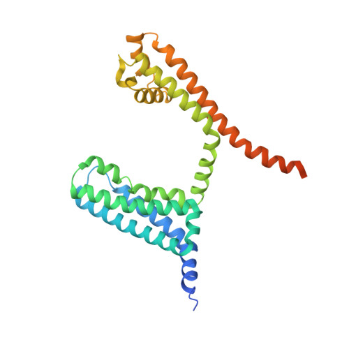
    5EK0

  • PubMed Abstract: 

    Voltage-gated sodium (Nav) channels propagate action potentials in excitable cells. Accordingly, Nav channels are therapeutic targets for many cardiovascular and neurological disorders. Selective inhibitors have been challenging to design because the nine mammalian Nav channel isoforms share high sequence identity and remain recalcitrant to high-resolution structural studies. Targeting the human Nav1.7 channel involved in pain perception, we present a protein-engineering strategy that has allowed us to determine crystal structures of a novel receptor site in complex with isoform-selective antagonists. GX-936 and related inhibitors bind to the activated state of voltage-sensor domain IV (VSD4), where their anionic aryl sulfonamide warhead engages the fourth arginine gating charge on the S4 helix. By opposing VSD4 deactivation, these compounds inhibit Nav1.7 through a voltage-sensor trapping mechanism, likely by stabilizing inactivated states of the channel. Residues from the S2 and S3 helices are key determinants of isoform selectivity, and bound phospholipids implicate the membrane as a modulator of channel function and pharmacology. Our results help to elucidate the molecular basis of voltage sensing and establish structural blueprints to design selective Nav channel antagonists.


  • Organizational Affiliation

    Department of Structural Biology, Genentech Inc., South San Francisco, CA 94080, USA.


Macromolecules
Find similar proteins by:  (by identity cutoff)  |  3D Structure
Entity ID: 1
MoleculeChains Sequence LengthOrganismDetailsImage
Chimera of bacterial Ion transport protein and human Sodium channel protein type 9 subunit alpha
A, B, C, D
296Aliarcobacter butzleri RM4018Homo sapiens
This entity is chimeric
Mutation(s): 0 
Gene Names: Abu_1752SCN9A
Membrane Entity: Yes 
UniProt & NIH Common Fund Data Resources
Find proteins for A8EVM5 (Aliarcobacter butzleri (strain RM4018))
Explore A8EVM5 
Go to UniProtKB:  A8EVM5
Find proteins for Q15858 (Homo sapiens)
Explore Q15858 
Go to UniProtKB:  Q15858
PHAROS:  Q15858
GTEx:  ENSG00000169432 
Entity Groups  
Sequence Clusters30% Identity50% Identity70% Identity90% Identity95% Identity100% Identity
UniProt GroupsA8EVM5Q15858
Sequence Annotations
Expand
  • Reference Sequence
Small Molecules
Ligands 2 Unique
IDChains Name / Formula / InChI Key2D Diagram3D Interactions
PX4
Query on PX4

Download Ideal Coordinates CCD File 
AA [auth D]
BA [auth D]
E [auth A]
F [auth A]
G [auth A]
AA [auth D],
BA [auth D],
E [auth A],
F [auth A],
G [auth A],
H [auth A],
I [auth A],
J [auth A],
K [auth A],
M [auth B],
N [auth B],
O [auth B],
P [auth B],
Q [auth B],
S [auth C],
T [auth C],
U [auth C],
V [auth C],
X [auth D],
Y [auth D],
Z [auth D]
1,2-DIMYRISTOYL-SN-GLYCERO-3-PHOSPHOCHOLINE
C36 H73 N O8 P
CITHEXJVPOWHKC-UUWRZZSWSA-O
5P2
Query on 5P2

Download Ideal Coordinates CCD File 
CA [auth D],
L [auth A],
R [auth B],
W [auth C]
3-cyano-4-[2-[2-(1-ethylazetidin-3-yl)pyrazol-3-yl]-4-(trifluoromethyl)phenoxy]-~{N}-(1,2,4-thiadiazol-5-yl)benzenesulfonamide
C24 H20 F3 N7 O3 S2
UCAVSLIXTXZSRD-UHFFFAOYSA-N
Experimental Data & Validation

Experimental Data

  • Method: X-RAY DIFFRACTION
  • Resolution: 3.53 Å
  • R-Value Free: 0.272 
  • R-Value Work: 0.243 
  • R-Value Observed: 0.244 
  • Space Group: C 2 2 21
Unit Cell:
Length ( Å )Angle ( ˚ )
a = 169.42α = 90
b = 188.83β = 90
c = 171.13γ = 90
Software Package:
Software NamePurpose
BUSTERrefinement
Aimlessdata scaling
PHENIXphasing
iMOSFLMdata reduction

Structure Validation

View Full Validation Report


Ligand Structure Quality Assessment 


Entry History 

Deposition Data

Revision History  (Full details and data files)

  • Version 1.0: 2015-12-23
    Type: Initial release
  • Version 1.1: 2015-12-30
    Changes: Database references
  • Version 1.2: 2023-09-27
    Changes: Data collection, Database references, Derived calculations, Refinement description