Ligand Binding Enhances Millisecond Conformational Exchange in Xylanase B2 from Streptomyces lividans.
Gagne, D., Narayanan, C., Nguyen-Thi, N., Roux, L.D., Bernard, D.N., Brunzelle, J.S., Couture, J.F., Agarwal, P.K., Doucet, N.(2016) Biochemistry 55: 4184-4196
- PubMed: 27387012
- DOI: https://doi.org/10.1021/acs.biochem.6b00130
- Primary Citation of Related Structures:
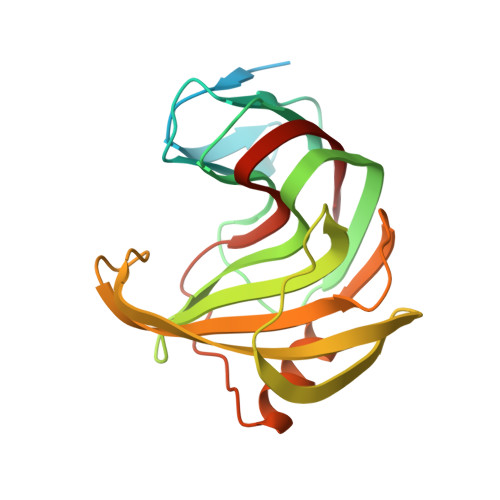
5EJ3 - PubMed Abstract:
Xylanases catalyze the hydrolysis of xylan, an abundant carbon and energy source with important commercial ramifications. Despite tremendous efforts devoted to the catalytic improvement of xylanases, success remains limited because of our relatively poor understanding of their molecular properties. Previous reports suggested the potential role of atomic-scale residue dynamics in modulating the catalytic activity of GH11 xylanases; however, dynamics in these studies was probed on time scales orders of magnitude faster than the catalytic time frame. Here, we used nuclear magnetic resonance titration and relaxation dispersion experiments ((15)N-CPMG) in combination with X-ray crystallography and computational simulations to probe conformational motions occurring on the catalytically relevant millisecond time frame in xylanase B2 (XlnB2) and its catalytically impaired mutant E87A from Streptomyces lividans 66. Our results show distinct dynamical properties for the apo and ligand-bound states of the enzymes. The apo form of XlnB2 experiences conformational exchange for residues in the fingers and palm regions of the catalytic cleft, while the catalytically impaired E87A variant displays millisecond dynamics only in the fingers, demonstrating the long-range effect of the mutation on flexibility. Ligand binding induces enhanced conformational exchange of residues interacting with the ligand in the fingers and thumb loop regions, emphasizing the potential role of residue motions in the fingers and thumb loop regions for recognition, positioning, processivity, and/or stabilization of ligands in XlnB2. To the best of our knowledge, this work represents the first experimental characterization of millisecond dynamics in a GH11 xylanase family member. These results offer new insights into the potential role of conformational exchange in GH11 enzymes, providing essential dynamic information to help improve protein engineering and design applications.
Organizational Affiliation:
INRS-Institut Armand-Frappier, Université du Québec , 531 Boul. des Prairies, Laval, Québec H7V 1B7, Canada.