5EIL

Computational design of a high-affinity metalloprotein homotrimer containing a metal chelating non-canonical amino acid


Experimental Data Snapshot

  • Method: X-RAY DIFFRACTION
  • Resolution: 2.25 Å
  • R-Value Free: 0.245 
  • R-Value Work: 0.214 
  • R-Value Observed: 0.217 

wwPDB Validation   3D Report Full Report


This is version 2.0 of the entry. See complete history


Literature

Computational design of a homotrimeric metalloprotein with a trisbipyridyl core.

Mills, J.H.Sheffler, W.Ener, M.E.Almhjell, P.J.Oberdorfer, G.Pereira, J.H.Parmeggiani, F.Sankaran, B.Zwart, P.H.Baker, D.

(2016) Proc Natl Acad Sci U S A 113: 15012-15017

  • DOI: https://doi.org/10.1073/pnas.1600188113
  • Primary Citation of Related Structures:  
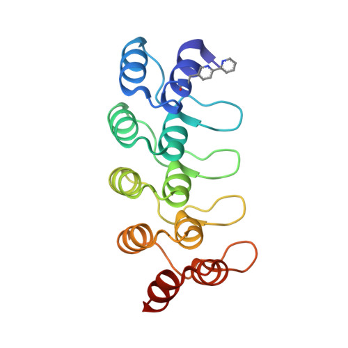
    5EIL

  • PubMed Abstract: 

    Metal-chelating heteroaryl small molecules have found widespread use as building blocks for coordination-driven, self-assembling nanostructures. The metal-chelating noncanonical amino acid (2,2'-bipyridin-5yl)alanine (Bpy-ala) could, in principle, be used to nucleate specific metalloprotein assemblies if introduced into proteins such that one assembly had much lower free energy than all alternatives. Here we describe the use of the Rosetta computational methodology to design a self-assembling homotrimeric protein with [Fe(Bpy-ala) 3 ] 2+ complexes at the interface between monomers. X-ray crystallographic analysis of the homotrimer showed that the design process had near-atomic-level accuracy: The all-atom rmsd between the design model and crystal structure for the residues at the protein interface is ∼1.4 Å. These results demonstrate that computational protein design together with genetically encoded noncanonical amino acids can be used to drive formation of precisely specified metal-mediated protein assemblies that could find use in a wide range of photophysical applications.


  • Organizational Affiliation

    Department of Biochemistry and the Institute for Protein Design, University of Washington, Seattle, WA 98195.


Macromolecules
Find similar proteins by:  (by identity cutoff)  |  3D Structure
Entity ID: 1
MoleculeChains Sequence LengthOrganismDetailsImage
TRI-05
A, B, C
159synthetic constructMutation(s): 0 
Entity Groups  
Sequence Clusters30% Identity50% Identity70% Identity90% Identity95% Identity100% Identity
Sequence Annotations
Expand
  • Reference Sequence
Small Molecules
Ligands 1 Unique
IDChains Name / Formula / InChI Key2D Diagram3D Interactions
FE
Query on FE

Download Ideal Coordinates CCD File 
D [auth A]FE (III) ION
Fe
VTLYFUHAOXGGBS-UHFFFAOYSA-N
Modified Residues  1 Unique
IDChains TypeFormula2D DiagramParent
BP5
Query on BP5
A, B, C
L-PEPTIDE LINKINGC13 H13 N3 O2ALA
Experimental Data & Validation

Experimental Data

  • Method: X-RAY DIFFRACTION
  • Resolution: 2.25 Å
  • R-Value Free: 0.245 
  • R-Value Work: 0.214 
  • R-Value Observed: 0.217 
  • Space Group: P 1 21 1
Unit Cell:
Length ( Å )Angle ( ˚ )
a = 63.107α = 90
b = 63.94β = 116.87
c = 64.831γ = 90
Software Package:
Software NamePurpose
PHENIXrefinement
xia2data scaling
XDSdata scaling
PHASERphasing

Structure Validation

View Full Validation Report


Entry History & Funding Information

Deposition Data


Funding OrganizationLocationGrant Number
National Institutes of Health/National Institute of General Medical Sciences (NIH/NIGMS)United StatesF32GM099210

Revision History  (Full details and data files)

  • Version 1.0: 2016-11-16
    Type: Initial release
  • Version 1.1: 2017-01-18
    Changes: Database references
  • Version 1.2: 2017-09-27
    Changes: Author supporting evidence, Refinement description
  • Version 1.3: 2019-12-25
    Changes: Author supporting evidence
  • Version 2.0: 2023-11-15
    Changes: Advisory, Atomic model, Data collection, Database references, Derived calculations