5EGP

Crystal structure of the S-methyltransferase TmtA


Experimental Data Snapshot

  • Method: X-RAY DIFFRACTION
  • Resolution: 1.50 Å
  • R-Value Free: 0.164 
  • R-Value Work: 0.132 
  • R-Value Observed: 0.134 

wwPDB Validation   3D Report Full Report


Ligand Structure Quality Assessment 


This is version 1.1 of the entry. See complete history


Literature

Sequential Inactivation of Gliotoxin by the S-Methyltransferase TmtA.

Duell, E.R.Glaser, M.Le Chapelain, C.Antes, I.Groll, M.Huber, E.M.

(2016) ACS Chem Biol 11: 1082-1089

  • DOI: https://doi.org/10.1021/acschembio.5b00905
  • Primary Citation of Related Structures:  
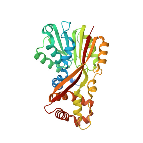
    5EGP

  • PubMed Abstract: 

    The epipolythiodioxopiperazine (ETP) gliotoxin mediates toxicity via its reactive thiol groups and thereby contributes to virulence of the human pathogenic fungus Aspergillus fumigatus. Self-intoxication of the mold is prevented either by reversible oxidation of reduced gliotoxin or by irreversible conversion to bis(methylthio)gliotoxin. The latter is produced by the S-methyltransferase TmtA and attenuates ETP biosynthesis. Here, we report the crystal structure of TmtA in complex with S-(5'-adenosyl)-l-homocysteine. TmtA features one substrate and one cofactor binding pocket per protein, and thus, bis-thiomethylation of gliotoxin occurs sequentially. Molecular docking of substrates and products into the active site of TmtA reveals that gliotoxin forms specific interactions with the protein surroundings, and free energy calculations indicate that methylation of the C10a-SH group precedes alkylation of the C3-SH site. Altogether, TmtA is well suited to selectively convert gliotoxin and to control its biosynthesis, suggesting that homologous enzymes serve to regulate the production of their toxic natural sulfur compounds in a similar manner.


  • Organizational Affiliation

    Center for Integrated Protein Science Munich (CIPSM) at the Department Chemie, Technische Universität München , Lichtenbergstr. 4, 85748 Garching, Germany.


Macromolecules
Find similar proteins by:  (by identity cutoff)  |  3D Structure
Entity ID: 1
MoleculeChains Sequence LengthOrganismDetailsImage
UbiE/COQ5 family methyltransferase, putative
A, B
314Aspergillus fumigatus Z5Mutation(s): 0 
Gene Names: Y699_02735
UniProt
Find proteins for Q4X158 (Aspergillus fumigatus (strain ATCC MYA-4609 / CBS 101355 / FGSC A1100 / Af293))
Explore Q4X158 
Go to UniProtKB:  Q4X158
Entity Groups  
Sequence Clusters30% Identity50% Identity70% Identity90% Identity95% Identity100% Identity
UniProt GroupQ4X158
Sequence Annotations
Expand
  • Reference Sequence
Small Molecules
Ligands 3 Unique
IDChains Name / Formula / InChI Key2D Diagram3D Interactions
SAH
Query on SAH

Download Ideal Coordinates CCD File 
C [auth A],
H [auth B]
S-ADENOSYL-L-HOMOCYSTEINE
C14 H20 N6 O5 S
ZJUKTBDSGOFHSH-WFMPWKQPSA-N
SO4
Query on SO4

Download Ideal Coordinates CCD File 
D [auth A],
I [auth B]
SULFATE ION
O4 S
QAOWNCQODCNURD-UHFFFAOYSA-L
ACT
Query on ACT

Download Ideal Coordinates CCD File 
E [auth A]
F [auth A]
G [auth A]
J [auth B]
K [auth B]
E [auth A],
F [auth A],
G [auth A],
J [auth B],
K [auth B],
L [auth B]
ACETATE ION
C2 H3 O2
QTBSBXVTEAMEQO-UHFFFAOYSA-M
Experimental Data & Validation

Experimental Data

  • Method: X-RAY DIFFRACTION
  • Resolution: 1.50 Å
  • R-Value Free: 0.164 
  • R-Value Work: 0.132 
  • R-Value Observed: 0.134 
  • Space Group: P 1 21 1
Unit Cell:
Length ( Å )Angle ( ˚ )
a = 49.1α = 90
b = 109.21β = 112.44
c = 59.94γ = 90
Software Package:
Software NamePurpose
REFMACrefinement
XDSdata reduction
XSCALEdata scaling
SHARPphasing

Structure Validation

View Full Validation Report


Ligand Structure Quality Assessment 


Entry History & Funding Information

Deposition Data


Funding OrganizationLocationGrant Number
DFGGermanySFB749
Hans-Fischer-GesellschaftGermany--

Revision History  (Full details and data files)

  • Version 1.0: 2016-02-03
    Type: Initial release
  • Version 1.1: 2016-04-27
    Changes: Database references