5ED4

Structure of a PhoP-DNA complex


Experimental Data Snapshot

  • Method: X-RAY DIFFRACTION
  • Resolution: 2.40 Å
  • R-Value Free: 0.227 
  • R-Value Work: 0.183 
  • R-Value Observed: 0.185 

wwPDB Validation   3D Report Full Report


This is version 1.3 of the entry. See complete history


Literature

Structural basis of DNA sequence recognition by the response regulator PhoP in Mycobacterium tuberculosis.

He, X.Wang, L.Wang, S.

(2016) Sci Rep 6: 24442-24442

  • DOI: https://doi.org/10.1038/srep24442
  • Primary Citation of Related Structures:  
    5ED4

  • PubMed Abstract: 

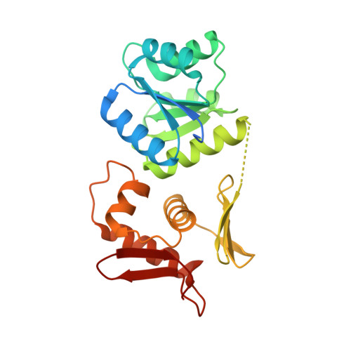
    The transcriptional regulator PhoP is an essential virulence factor in Mycobacterium tuberculosis, and it presents a target for the development of new anti-tuberculosis drugs and attenuated tuberculosis vaccine strains. PhoP binds to DNA as a highly cooperative dimer by recognizing direct repeats of 7-bp motifs with a 4-bp spacer. To elucidate the PhoP-DNA binding mechanism, we determined the crystal structure of the PhoP-DNA complex. The structure revealed a tandem PhoP dimer that bound to the direct repeat. The surprising tandem arrangement of the receiver domains allowed the four domains of the PhoP dimer to form a compact structure, accounting for the strict requirement of a 4-bp spacer and the highly cooperative binding of the dimer. The PhoP-DNA interactions exclusively involved the effector domain. The sequence-recognition helix made contact with the bases of the 7-bp motif in the major groove, and the wing interacted with the adjacent minor groove. The structure provides a starting point for the elucidation of the mechanism by which PhoP regulates the virulence of M. tuberculosis and guides the design of screening platforms for PhoP inhibitors.


  • Organizational Affiliation

    Department of Biochemistry &Molecular Biology, Uniformed Services University of the Health Sciences, 4301 Jones Bridge Road, Bethesda, Maryland 20814, USA.


Macromolecules

Find similar proteins by:  (by identity cutoff)  |  3D Structure
Entity ID: 1
MoleculeChains Sequence LengthOrganismDetailsImage
Response regulator
A, B, E, F
250Mycobacterium tuberculosisMutation(s): 0 
Gene Names: 
EC: 3.1.3.1
UniProt
Find proteins for P71814 (Mycobacterium tuberculosis (strain ATCC 25618 / H37Rv))
Explore P71814 
Go to UniProtKB:  P71814
Entity Groups  
Sequence Clusters30% Identity50% Identity70% Identity90% Identity95% Identity100% Identity
UniProt GroupP71814
Sequence Annotations
Expand
  • Reference Sequence
Find similar nucleic acids by:  (by identity cutoff)  |  3D Structure
Entity ID: 2
MoleculeChains LengthOrganismImage
DNA (26-MER)
C, G
26synthetic construct
Sequence Annotations
Expand
  • Reference Sequence
Find similar nucleic acids by:  (by identity cutoff)  |  3D Structure
Entity ID: 3
MoleculeChains LengthOrganismImage
DNA (26-MER)
D, H
26synthetic construct
Sequence Annotations
Expand
  • Reference Sequence
Small Molecules
Ligands 3 Unique
IDChains Name / Formula / InChI Key2D Diagram3D Interactions
CAC
Query on CAC

Download Ideal Coordinates CCD File 
Q [auth E]CACODYLATE ION
C2 H6 As O2
OGGXGZAMXPVRFZ-UHFFFAOYSA-M
EDO
Query on EDO

Download Ideal Coordinates CCD File 
J [auth A],
K [auth A],
N [auth B],
P [auth E]
1,2-ETHANEDIOL
C2 H6 O2
LYCAIKOWRPUZTN-UHFFFAOYSA-N
CA
Query on CA

Download Ideal Coordinates CCD File 
I [auth A]
L [auth B]
M [auth B]
O [auth E]
R [auth F]
I [auth A],
L [auth B],
M [auth B],
O [auth E],
R [auth F],
S [auth F]
CALCIUM ION
Ca
BHPQYMZQTOCNFJ-UHFFFAOYSA-N
Experimental Data & Validation

Experimental Data

  • Method: X-RAY DIFFRACTION
  • Resolution: 2.40 Å
  • R-Value Free: 0.227 
  • R-Value Work: 0.183 
  • R-Value Observed: 0.185 
  • Space Group: P 21 21 21
Unit Cell:
Length ( Å )Angle ( ˚ )
a = 92.24α = 90
b = 98.176β = 90
c = 167.888γ = 90
Software Package:
Software NamePurpose
HKL-2000data scaling
BUSTER-TNTrefinement
PDB_EXTRACTdata extraction
PHASERphasing
Cootmodel building
HKL-2000data reduction

Structure Validation

View Full Validation Report


Entry History & Funding Information

Deposition Data

  • Released Date: 2016-04-27 
  • Deposition Author(s): Wang, S.

Funding OrganizationLocationGrant Number
National Institutes of Health/National Institute of General Medical Sciences (NIH/NIGMS)United StatesR01GM079185

Revision History  (Full details and data files)

  • Version 1.0: 2016-04-27
    Type: Initial release
  • Version 1.1: 2017-09-13
    Changes: Author supporting evidence, Derived calculations
  • Version 1.2: 2019-12-25
    Changes: Author supporting evidence
  • Version 1.3: 2023-09-27
    Changes: Data collection, Database references, Derived calculations, Refinement description