5E9Z

Cytochrome P450 BM3 mutant M11


Experimental Data Snapshot

  • Method: X-RAY DIFFRACTION
  • Resolution: 2.23 Å
  • R-Value Free: 0.225 
  • R-Value Work: 0.188 
  • R-Value Observed: 0.190 

wwPDB Validation   3D Report Full Report


Ligand Structure Quality Assessment 


This is version 1.2 of the entry. See complete history


Literature

Insights into regioselective metabolism of mefenamic acid by cytochrome P450 BM3 mutants through crystallography, docking, molecular dynamics, and free energy calculations.

Capoferri, L.Leth, R.Ter Haar, E.Mohanty, A.K.Grootenhuis, P.D.Vottero, E.Commandeur, J.N.Vermeulen, N.P.Jrgensen, F.S.Olsen, L.Geerke, D.P.

(2016) Proteins 84: 383-396

  • DOI: https://doi.org/10.1002/prot.24985
  • Primary Citation of Related Structures:  
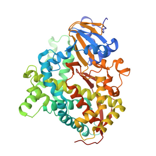
    5E9Z

  • PubMed Abstract: 

    Cytochrome P450 BM3 (CYP102A1) mutant M11 is able to metabolize a wide range of drugs and drug-like compounds. Among these, M11 was recently found to be able to catalyze formation of human metabolites of mefenamic acid and other nonsteroidal anti-inflammatory drugs (NSAIDs). Interestingly, single active-site mutations such as V87I were reported to invert regioselectivity in NSAID hydroxylation. In this work, we combine crystallography and molecular simulation to study the effect of single mutations on binding and regioselective metabolism of mefenamic acid by M11 mutants. The heme domain of the protein mutant M11 was expressed, purified, and crystallized, and its X-ray structure was used as template for modeling. A multistep approach was used that combines molecular docking, molecular dynamics (MD) simulation, and binding free-energy calculations to address protein flexibility. In this way, preferred binding modes that are consistent with oxidation at the experimentally observed sites of metabolism (SOMs) were identified. Whereas docking could not be used to retrospectively predict experimental trends in regioselectivity, we were able to rank binding modes in line with the preferred SOMs of mefenamic acid by M11 and its mutants by including protein flexibility and dynamics in free-energy computation. In addition, we could obtain structural insights into the change in regioselectivity of mefenamic acid hydroxylation due to single active-site mutations. Our findings confirm that use of MD and binding free-energy calculation is useful for studying biocatalysis in those cases in which enzyme binding is a critical event in determining the selective metabolism of a substrate.


  • Organizational Affiliation

    AIMMS Division of Molecular Toxicology, Department of Chemistry and Pharmaceutical Sciences, VU University, De Boelelaan 1083, 1081 HV Amsterdam, the Netherlands.


Macromolecules
Find similar proteins by:  (by identity cutoff)  |  3D Structure
Entity ID: 1
MoleculeChains Sequence LengthOrganismDetailsImage
Bifunctional cytochrome P450/NADPH--P450 reductase
A, B, C, D
469Priestia megateriumMutation(s): 10 
Gene Names: cyp102A1cyp102
EC: 1.14.14.1 (PDB Primary Data), 1.6.2.4 (PDB Primary Data)
UniProt
Find proteins for P14779 (Priestia megaterium (strain ATCC 14581 / DSM 32 / CCUG 1817 / JCM 2506 / NBRC 15308 / NCIMB 9376 / NCTC 10342 / NRRL B-14308 / VKM B-512 / Ford 19))
Explore P14779 
Go to UniProtKB:  P14779
Entity Groups  
Sequence Clusters30% Identity50% Identity70% Identity90% Identity95% Identity100% Identity
UniProt GroupP14779
Sequence Annotations
Expand
  • Reference Sequence
Small Molecules
Ligands 3 Unique
IDChains Name / Formula / InChI Key2D Diagram3D Interactions
PP9
Query on PP9

Download Ideal Coordinates CCD File 
F [auth A],
H [auth B],
J [auth C],
L [auth D]
PROTOPORPHYRIN IX
C34 H34 N4 O4
FEDYMSUPMFCVOD-UJJXFSCMSA-N
DTT
Query on DTT

Download Ideal Coordinates CCD File 
G [auth B]2,3-DIHYDROXY-1,4-DITHIOBUTANE
C4 H10 O2 S2
VHJLVAABSRFDPM-IMJSIDKUSA-N
FE2
Query on FE2

Download Ideal Coordinates CCD File 
E [auth A],
I [auth B],
K [auth C],
M [auth D]
FE (II) ION
Fe
CWYNVVGOOAEACU-UHFFFAOYSA-N
Experimental Data & Validation

Experimental Data

  • Method: X-RAY DIFFRACTION
  • Resolution: 2.23 Å
  • R-Value Free: 0.225 
  • R-Value Work: 0.188 
  • R-Value Observed: 0.190 
  • Space Group: C 1 2 1
Unit Cell:
Length ( Å )Angle ( ˚ )
a = 379.122α = 90
b = 59.72β = 95.67
c = 95.587γ = 90
Software Package:
Software NamePurpose
BUSTER-TNTrefinement
SCALAdata scaling
PHASERphasing
Cootmodel building
PDB_EXTRACTdata extraction

Structure Validation

View Full Validation Report


Ligand Structure Quality Assessment 


Entry History 

Revision History  (Full details and data files)

  • Version 1.0: 2016-01-27
    Type: Initial release
  • Version 1.1: 2016-02-24
    Changes: Database references
  • Version 1.2: 2023-09-27
    Changes: Data collection, Database references, Derived calculations, Refinement description