5E78

Crystal structure of P450 BM3 heme domain variant complexed with Co(III)Sep


Experimental Data Snapshot

  • Method: X-RAY DIFFRACTION
  • Resolution: 2.00 Å
  • R-Value Free: 0.212 
  • R-Value Work: 0.175 
  • R-Value Observed: 0.177 

wwPDB Validation   3D Report Full Report


Ligand Structure Quality Assessment 


This is version 1.4 of the entry. See complete history


Literature

Crystallographic insights into a cobalt (III) sepulchrate based alternative cofactor system of P450 BM3 monooxygenase.

Panneerselvam, S.Shehzad, A.Mueller-Dieckmann, J.Wilmanns, M.Bocola, M.Davari, M.D.Schwaneberg, U.

(2018) Biochim Biophys Acta 1866: 134-140

  • DOI: https://doi.org/10.1016/j.bbapap.2017.07.010
  • Primary Citation of Related Structures:  
    5E78

  • PubMed Abstract: 

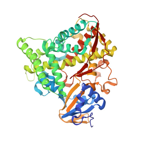
    P450 BM3 is a multi-domain heme-containing soluble bacterial monooxygenase. P450 BM3 and variants are known to oxidize structurally diverse substrates. Crystal structures of individual domains of P450 BM3 are available. However, the spatial organization of the full-length protein is unknown. In this study, crystal structures of the P450 BM3 M7 heme domain variant with and without cobalt (III) sepulchrate are reported. Cobalt (III) sepulchrate acts as an electron shuttle in an alternative cofactor system employing zinc dust as the electron source. The crystal structure shows a binding site for the mediator cobalt (III) sepulchrate at the entrance of the substrate access channel. The mediator occupies an unusual position which is far from the active site and distinct from the binding of the natural redox partner (FAD/NADPH binding domain).


  • Organizational Affiliation

    HASYLAB, DESY, Notkestrasse 85, 22603 Hamburg, Germany.


Macromolecules
Find similar proteins by:  (by identity cutoff)  |  3D Structure
Entity ID: 1
MoleculeChains Sequence LengthOrganismDetailsImage
Bifunctional P-450/NADPH-P450 reductase
A, B
455Priestia megateriumMutation(s): 3 
Gene Names: cyp102A1cyp102
EC: 1.14.14.1 (PDB Primary Data), 1.6.2.4 (PDB Primary Data)
UniProt
Find proteins for P14779 (Priestia megaterium (strain ATCC 14581 / DSM 32 / CCUG 1817 / JCM 2506 / NBRC 15308 / NCIMB 9376 / NCTC 10342 / NRRL B-14308 / VKM B-512 / Ford 19))
Explore P14779 
Go to UniProtKB:  P14779
Entity Groups  
Sequence Clusters30% Identity50% Identity70% Identity90% Identity95% Identity100% Identity
UniProt GroupP14779
Sequence Annotations
Expand
  • Reference Sequence
Experimental Data & Validation

Experimental Data

  • Method: X-RAY DIFFRACTION
  • Resolution: 2.00 Å
  • R-Value Free: 0.212 
  • R-Value Work: 0.175 
  • R-Value Observed: 0.177 
  • Space Group: P 21 21 21
Unit Cell:
Length ( Å )Angle ( ˚ )
a = 58.97α = 90
b = 128.54β = 90
c = 150.06γ = 90
Software Package:
Software NamePurpose
REFMACrefinement
XDSdata reduction
XSCALEdata scaling
PHASESphasing

Structure Validation

View Full Validation Report


Ligand Structure Quality Assessment 


Entry History 

Deposition Data

Revision History  (Full details and data files)

  • Version 1.0: 2016-09-28
    Type: Initial release
  • Version 1.1: 2017-11-22
    Changes: Database references
  • Version 1.2: 2017-12-06
    Changes: Database references
  • Version 1.3: 2019-07-17
    Changes: Data collection
  • Version 1.4: 2024-01-10
    Changes: Data collection, Database references, Derived calculations, Refinement description