5E4I

Crystal structure of mouse CNTN5 Ig1-Ig4 domains


Experimental Data Snapshot

  • Method: X-RAY DIFFRACTION
  • Resolution: 2.60 Å
  • R-Value Free: 0.264 
  • R-Value Work: 0.195 
  • R-Value Observed: 0.201 

wwPDB Validation   3D Report Full Report


Ligand Structure Quality Assessment 


This is version 1.5 of the entry. See complete history


Literature

Structural Basis for Interactions Between Contactin Family Members and Protein-tyrosine Phosphatase Receptor Type G in Neural Tissues.

Nikolaienko, R.M.Hammel, M.Dubreuil, V.Zalmai, R.Hall, D.R.Mehzabeen, N.Karuppan, S.J.Harroch, S.Stella, S.L.Bouyain, S.

(2016) J Biol Chem 291: 21335-21349

  • DOI: https://doi.org/10.1074/jbc.M116.742163
  • Primary Citation of Related Structures:  
    5E4I, 5E4Q, 5E4S, 5E52, 5E53, 5E55, 5E5R, 5E5U, 5E7L, 5I99

  • PubMed Abstract: 

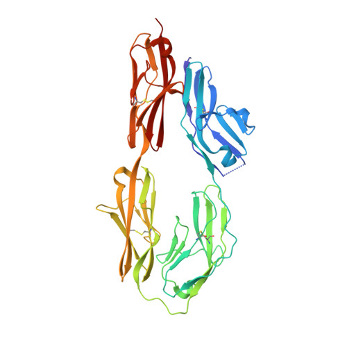
    Protein-tyrosine phosphatase receptor type G (RPTPγ/PTPRG) interacts in vitro with contactin-3-6 (CNTN3-6), a group of glycophosphatidylinositol-anchored cell adhesion molecules involved in the wiring of the nervous system. In addition to PTPRG, CNTNs associate with multiple transmembrane proteins and signal inside the cell via cis-binding partners to alleviate the absence of an intracellular region. Here, we use comprehensive biochemical and structural analyses to demonstrate that PTPRG·CNTN3-6 complexes share similar binding affinities and a conserved arrangement. Furthermore, as a first step to identifying PTPRG·CNTN complexes in vivo, we found that PTPRG and CNTN3 associate in the outer segments of mouse rod photoreceptor cells. In particular, PTPRG and CNTN3 form cis-complexes at the surface of photoreceptors yet interact in trans when expressed on the surfaces of apposing cells. Further structural analyses suggest that all CNTN ectodomains adopt a bent conformation and might lie parallel to the cell surface to accommodate these cis and trans binding modes. Taken together, these studies identify a PTPRG·CNTN complex in vivo and provide novel insights into PTPRG- and CNTN-mediated signaling.


  • Organizational Affiliation

    From the Division of Molecular Biology and Biochemistry, School of Biological Sciences, University of Missouri-Kansas City, Kansas City, Missouri 64110.


Macromolecules
Find similar proteins by:  (by identity cutoff)  |  3D Structure
Entity ID: 1
MoleculeChains Sequence LengthOrganismDetailsImage
Contactin-5384Mus musculusMutation(s): 0 
Gene Names: Cntn5
UniProt & NIH Common Fund Data Resources
Find proteins for P68500 (Mus musculus)
Explore P68500 
Go to UniProtKB:  P68500
IMPC:  MGI:3042287
Entity Groups  
Sequence Clusters30% Identity50% Identity70% Identity90% Identity95% Identity100% Identity
UniProt GroupP68500
Sequence Annotations
Expand
  • Reference Sequence
Small Molecules
Ligands 1 Unique
IDChains Name / Formula / InChI Key2D Diagram3D Interactions
NAG
Query on NAG

Download Ideal Coordinates CCD File 
B [auth A],
C [auth A]
2-acetamido-2-deoxy-beta-D-glucopyranose
C8 H15 N O6
OVRNDRQMDRJTHS-FMDGEEDCSA-N
Experimental Data & Validation

Experimental Data

  • Method: X-RAY DIFFRACTION
  • Resolution: 2.60 Å
  • R-Value Free: 0.264 
  • R-Value Work: 0.195 
  • R-Value Observed: 0.201 
  • Space Group: C 1 2 1
Unit Cell:
Length ( Å )Angle ( ˚ )
a = 179.975α = 90
b = 50.659β = 101.67
c = 51.015γ = 90
Software Package:
Software NamePurpose
HKL-2000data collection
HKL-2000data scaling
HKL-2000data reduction
PHENIXrefinement
Cootmodel building
PDB_EXTRACTdata extraction
PHASERphasing

Structure Validation

View Full Validation Report


Ligand Structure Quality Assessment 


Entry History & Funding Information

Deposition Data


Funding OrganizationLocationGrant Number
National Institutes of Health/National Institute of General Medical Sciences (NIH/NIGMS)United States--

Revision History  (Full details and data files)

  • Version 1.0: 2016-08-31
    Type: Initial release
  • Version 1.1: 2016-11-23
    Changes: Database references
  • Version 1.2: 2017-09-27
    Changes: Author supporting evidence, Database references
  • Version 1.3: 2019-12-25
    Changes: Author supporting evidence
  • Version 1.4: 2020-07-29
    Type: Remediation
    Reason: Carbohydrate remediation
    Changes: Data collection, Derived calculations, Structure summary
  • Version 1.5: 2023-09-27
    Changes: Data collection, Database references, Refinement description, Structure summary