5E1R

Crystal structure of pecan (carya illinoinensis) vicilin, a new food allergen


Experimental Data Snapshot

  • Method: X-RAY DIFFRACTION
  • Resolution: 2.65 Å
  • R-Value Free: 0.262 
  • R-Value Work: 0.218 
  • R-Value Observed: 0.219 

wwPDB Validation   3D Report Full Report


This is version 1.1 of the entry. See complete history


Literature

Identification and Characterization of a New Pecan [Carya illinoinensis (Wangenh.) K. Koch] Allergen, Car i 2.

Zhang, Y.Lee, B.Du, W.X.Lyu, S.C.Nadeau, K.C.Grauke, L.J.Zhang, Y.Wang, S.Fan, Y.Yi, J.McHugh, T.H.

(2016) J Agric Food Chem 64: 4146-4151

  • DOI: https://doi.org/10.1021/acs.jafc.6b00884
  • Primary Citation of Related Structures:  
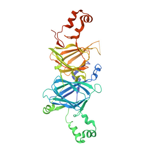
    5E1R

  • PubMed Abstract: 

    The 7S vicilin and 11S legumin seed storage globulins belong to the cupin protein superfamily and are major food allergens in many foods from the "big eight" food allergen groups. Here, for the first time, pecan vicilin was found to be a food allergen. Western blot experiments revealed that 30% of 27 sera used in this study and 24% of the sera from 25 patients with double-blind, placebo controlled clinical pecan allergy contained IgE antibodies specific to pecan vicilin. This allergen consists of a low-complexity region at its N-terminal and a structured domain at the C-terminal that contains two cupin motifs and forms homotrimers. The crystal structure of recombinant pecan vicilin was determined. The refined structure gave R/Rfree values of 0.218/0.262 for all data to 2.65 Å. There were two trimeric biological units in the crystallographic asymmetric unit. Pecan vicilin is also a copper protein. These data may facilitate the understanding of the nutritional value and the allergenicity relevance of the copper binding property of seed storage proteins in tree nuts.


  • Organizational Affiliation

    Western Regional Research Center, Pacific West Area, Agricultural Research Service, U.S. Department of Agriculture , 800 Buchanan Street, Albany, California 94710, United States.


Macromolecules
Find similar proteins by:  (by identity cutoff)  |  3D Structure
Entity ID: 1
MoleculeChains Sequence LengthOrganismDetailsImage
7S vicilin
A, B, C, D, E
A, B, C, D, E, F
426Carya illinoinensisMutation(s): 0 
Gene Names: pec2a1apec3a1apec4a1a
UniProt
Find proteins for B3STU4 (Carya illinoinensis)
Explore B3STU4 
Go to UniProtKB:  B3STU4
Entity Groups  
Sequence Clusters30% Identity50% Identity70% Identity90% Identity95% Identity100% Identity
UniProt GroupB3STU4
Sequence Annotations
Expand
  • Reference Sequence
Small Molecules
Ligands 2 Unique
IDChains Name / Formula / InChI Key2D Diagram3D Interactions
MPD
Query on MPD

Download Ideal Coordinates CCD File 
H [auth A]
J [auth B]
L [auth C]
N [auth D]
P [auth E]
H [auth A],
J [auth B],
L [auth C],
N [auth D],
P [auth E],
R [auth F]
(4S)-2-METHYL-2,4-PENTANEDIOL
C6 H14 O2
SVTBMSDMJJWYQN-YFKPBYRVSA-N
CU
Query on CU

Download Ideal Coordinates CCD File 
G [auth A]
I [auth B]
K [auth C]
M [auth D]
O [auth E]
G [auth A],
I [auth B],
K [auth C],
M [auth D],
O [auth E],
Q [auth F]
COPPER (II) ION
Cu
JPVYNHNXODAKFH-UHFFFAOYSA-N
Experimental Data & Validation

Experimental Data

  • Method: X-RAY DIFFRACTION
  • Resolution: 2.65 Å
  • R-Value Free: 0.262 
  • R-Value Work: 0.218 
  • R-Value Observed: 0.219 
  • Space Group: P 21 21 21
Unit Cell:
Length ( Å )Angle ( ˚ )
a = 42.691α = 90
b = 151.61β = 90
c = 342.782γ = 90
Software Package:
Software NamePurpose
HKL-2000data scaling
PHENIXrefinement
PDB_EXTRACTdata extraction
HKL-2000data reduction
PHASERphasing

Structure Validation

View Full Validation Report


Entry History 

Deposition Data

  • Released Date: 2016-08-10 
  • Deposition Author(s): Zhang, Y.Z.

Revision History  (Full details and data files)

  • Version 1.0: 2016-08-10
    Type: Initial release
  • Version 1.1: 2024-03-06
    Changes: Data collection, Database references, Derived calculations