5DZ2

Geosmin synthase from Streptomyces coelicolor N-terminal domain complexed with three Mg2+ ions and alendronic acid


Experimental Data Snapshot

  • Method: X-RAY DIFFRACTION
  • Resolution: 2.11 Å
  • R-Value Free: 0.250 
  • R-Value Work: 0.207 
  • R-Value Observed: 0.210 

wwPDB Validation   3D Report Full Report


Ligand Structure Quality Assessment 


This is version 1.4 of the entry. See complete history


Literature

Structural Studies of Geosmin Synthase, a Bifunctional Sesquiterpene Synthase with alpha alpha Domain Architecture That Catalyzes a Unique Cyclization-Fragmentation Reaction Sequence.

Harris, G.G.Lombardi, P.M.Pemberton, T.A.Matsui, T.Weiss, T.M.Cole, K.E.Koksal, M.Murphy, F.V.Vedula, L.S.Chou, W.K.Cane, D.E.Christianson, D.W.

(2015) Biochemistry 54: 7142-7155

  • DOI: https://doi.org/10.1021/acs.biochem.5b01143
  • Primary Citation of Related Structures:  
    5DW7, 5DZ2

  • PubMed Abstract: 

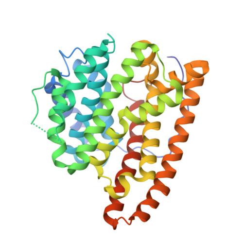
    Geosmin synthase from Streptomyces coelicolor (ScGS) catalyzes an unusual, metal-dependent terpenoid cyclization and fragmentation reaction sequence. Two distinct active sites are required for catalysis: the N-terminal domain catalyzes the ionization and cyclization of farnesyl diphosphate to form germacradienol and inorganic pyrophosphate (PPi), and the C-terminal domain catalyzes the protonation, cyclization, and fragmentation of germacradienol to form geosmin and acetone through a retro-Prins reaction. A unique αα domain architecture is predicted for ScGS based on amino acid sequence: each domain contains the metal-binding motifs typical of a class I terpenoid cyclase, and each domain requires Mg(2+) for catalysis. Here, we report the X-ray crystal structure of the unliganded N-terminal domain of ScGS and the structure of its complex with three Mg(2+) ions and alendronate. These structures highlight conformational changes required for active site closure and catalysis. Although neither full-length ScGS nor constructs of the C-terminal domain could be crystallized, homology models of the C-terminal domain were constructed on the basis of ∼36% sequence identity with the N-terminal domain. Small-angle X-ray scattering experiments yield low-resolution molecular envelopes into which the N-terminal domain crystal structure and the C-terminal domain homology model were fit, suggesting possible αα domain architectures as frameworks for bifunctional catalysis.


  • Organizational Affiliation

    Roy and Diana Vagelos Laboratories, Department of Chemistry, University of Pennsylvania , Philadelphia, Pennsylvania 19104-6323, United States.


Macromolecules
Find similar proteins by:  (by identity cutoff)  |  3D Structure
Entity ID: 1
MoleculeChains Sequence LengthOrganismDetailsImage
Germacradienol/geosmin synthase
A, B
338Streptomyces coelicolor A3(2)Mutation(s): 0 
Gene Names: cyc2SCO6073SC9B1.20
EC: 4.2.3.22 (PDB Primary Data), 4.2.3.75 (PDB Primary Data), 4.1.99.16 (PDB Primary Data)
UniProt
Find proteins for Q9X839 (Streptomyces coelicolor (strain ATCC BAA-471 / A3(2) / M145))
Explore Q9X839 
Go to UniProtKB:  Q9X839
Entity Groups  
Sequence Clusters30% Identity50% Identity70% Identity90% Identity95% Identity100% Identity
UniProt GroupQ9X839
Sequence Annotations
Expand
  • Reference Sequence
Experimental Data & Validation

Experimental Data

  • Method: X-RAY DIFFRACTION
  • Resolution: 2.11 Å
  • R-Value Free: 0.250 
  • R-Value Work: 0.207 
  • R-Value Observed: 0.210 
  • Space Group: P 43 21 2
Unit Cell:
Length ( Å )Angle ( ˚ )
a = 67.213α = 90
b = 67.213β = 90
c = 345.388γ = 90
Software Package:
Software NamePurpose
PHENIXrefinement
HKL-2000data reduction
HKL-2000data scaling
PHENIXphasing

Structure Validation

View Full Validation Report


Ligand Structure Quality Assessment 


Entry History & Funding Information

Deposition Data


Funding OrganizationLocationGrant Number
National Institutes of Health/National Institute of General Medical Sciences (NIH/NIGMS)United StatesGM56838

Revision History  (Full details and data files)

  • Version 1.0: 2015-12-09
    Type: Initial release
  • Version 1.1: 2015-12-23
    Changes: Database references
  • Version 1.2: 2017-09-13
    Changes: Author supporting evidence, Database references, Derived calculations
  • Version 1.3: 2019-12-25
    Changes: Author supporting evidence
  • Version 1.4: 2023-09-27
    Changes: Data collection, Database references, Derived calculations, Refinement description