5DXV

Crystal structure of Rethreaded DHFR


Experimental Data Snapshot

  • Method: X-RAY DIFFRACTION
  • Resolution: 1.55 Å
  • R-Value Free: 0.214 
  • R-Value Work: 0.185 
  • R-Value Observed: 0.186 

wwPDB Validation   3D Report Full Report


Ligand Structure Quality Assessment 


This is version 1.0 of the entry. See complete history


Literature

Protein rethreading: A novel approach to protein design.

Agah, S.Poulos, S.Yu, A.Kucharska, I.Faham, S.

(2016) Sci Rep 6: 26847-26847

  • DOI: https://doi.org/10.1038/srep26847
  • Primary Citation of Related Structures:  
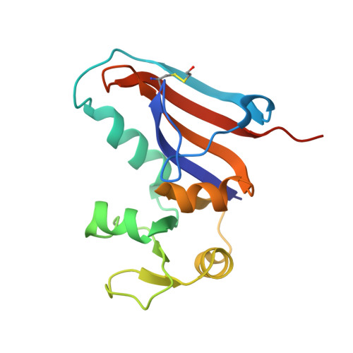
    5DXV

  • PubMed Abstract: 

    Protein engineering is an important tool for the design of proteins with novel and desirable features. Templates from the protein databank (PDB) are often used as initial models that can be modified to introduce new properties. We examine whether it is possible to reconnect a protein in a manner that generates a new topology yet preserves its structural integrity. Here, we describe the rethreading of dihydrofolate reductase (DHFR) from E. coli (wtDHFR). The rethreading process involved the removal of three native loops, and the introduction of three new loops with alternate connections. The structure of the rethreaded DHFR (rDHFR-1) was determined to 1.6 Å, demonstrating the success of the rethreading process. Both wtDHFR and rDHFR-1 exhibited similar affinities towards methotrexate. However, rDHFR-1 showed no reducing activity towards dihydrofolate, and exhibited about ~6-fold lower affinity towards NADPH than wtDHFR. This work demonstrates that protein rethreading can be a powerful tool for the design of a large array of proteins with novel structures and topologies, and that by careful rearrangement of a protein sequence, the sequence to structure relationship can be expanded substantially.


  • Organizational Affiliation

    Department of Molecular Physiology and Biological Physics, University of Virginia School of Medicine, Charlottesville, Virginia, 22903, United States.


Macromolecules
Find similar proteins by:  (by identity cutoff)  |  3D Structure
Entity ID: 1
MoleculeChains Sequence LengthOrganismDetailsImage
rethreaded DHFR
A, B
156synthetic constructMutation(s): 0 
Entity Groups  
Sequence Clusters30% Identity50% Identity70% Identity90% Identity95% Identity100% Identity
Sequence Annotations
Expand
  • Reference Sequence
Small Molecules
Ligands 4 Unique
IDChains Name / Formula / InChI Key2D Diagram3D Interactions
NAP
Query on NAP

Download Ideal Coordinates CCD File 
C [auth A]NADP NICOTINAMIDE-ADENINE-DINUCLEOTIDE PHOSPHATE
C21 H28 N7 O17 P3
XJLXINKUBYWONI-NNYOXOHSSA-N
PEG
Query on PEG

Download Ideal Coordinates CCD File 
D [auth A],
G [auth B]
DI(HYDROXYETHYL)ETHER
C4 H10 O3
MTHSVFCYNBDYFN-UHFFFAOYSA-N
EDO
Query on EDO

Download Ideal Coordinates CCD File 
E [auth A],
H [auth B]
1,2-ETHANEDIOL
C2 H6 O2
LYCAIKOWRPUZTN-UHFFFAOYSA-N
CL
Query on CL

Download Ideal Coordinates CCD File 
F [auth A],
I [auth B]
CHLORIDE ION
Cl
VEXZGXHMUGYJMC-UHFFFAOYSA-M
Modified Residues  1 Unique
IDChains TypeFormula2D DiagramParent
CME
Query on CME
A, B
L-PEPTIDE LINKINGC5 H11 N O3 S2CYS
Experimental Data & Validation

Experimental Data

  • Method: X-RAY DIFFRACTION
  • Resolution: 1.55 Å
  • R-Value Free: 0.214 
  • R-Value Work: 0.185 
  • R-Value Observed: 0.186 
  • Space Group: C 2 2 21
Unit Cell:
Length ( Å )Angle ( ˚ )
a = 44.817α = 90
b = 120.054β = 90
c = 111.187γ = 90
Software Package:
Software NamePurpose
REFMACrefinement
HKL-2000data reduction
SCALEPACKdata scaling
PHASERphasing

Structure Validation

View Full Validation Report


Ligand Structure Quality Assessment 


Entry History 

Deposition Data

  • Released Date: 2016-06-08 
  • Deposition Author(s): Faham, S.

Revision History  (Full details and data files)

  • Version 1.0: 2016-06-08
    Type: Initial release